. 2013 Apr;24(7):982-94.
doi: 10.1091/mbc.E12-09-0685. Epub 2013 Feb 27.

Visualization of actin filaments and monomers in somatic cell nuclei

Affiliations

Visualization of actin filaments and monomers in somatic cell nuclei

Brittany J Belin et al. Mol Biol Cell. 2013 Apr.

Abstract

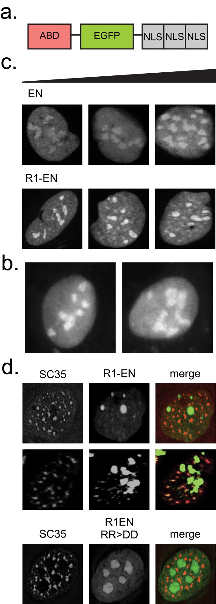
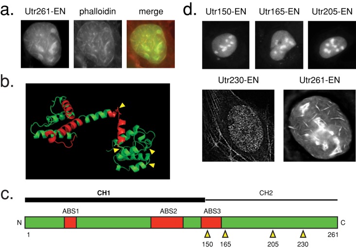
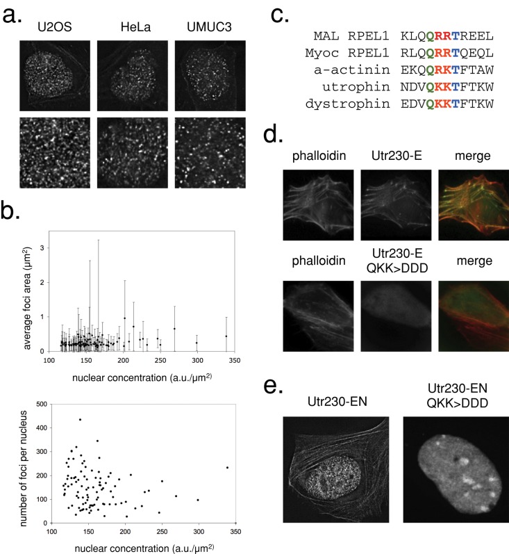
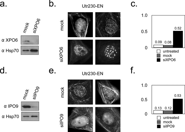
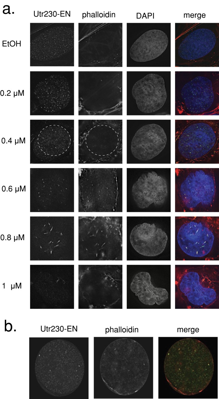
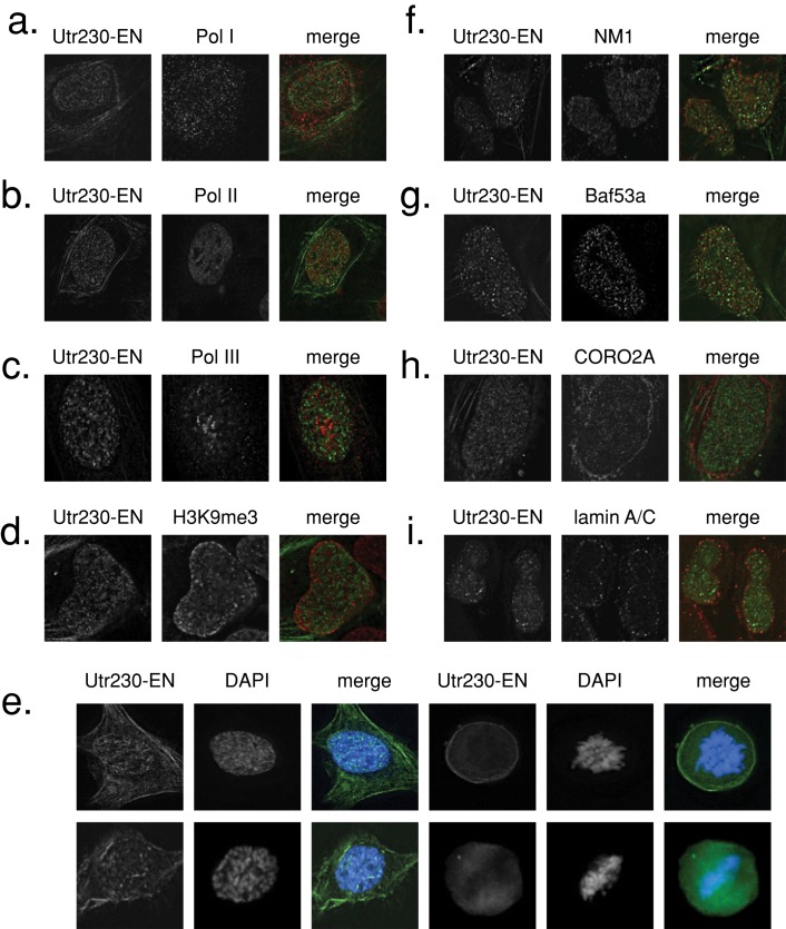
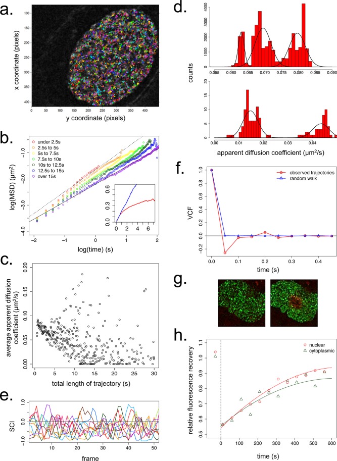
In addition to its long-studied presence in the cytoplasm, actin is also found in the nuclei of eukaryotic cells. The function and form (monomer, filament, or noncanonical oligomer) of nuclear actin are hotly debated, and its localization and dynamics are largely unknown. To determine the distribution of nuclear actin in live somatic cells and evaluate its potential functions, we constructed and validated fluorescent nuclear actin probes. Monomeric actin probes concentrate in nuclear speckles, suggesting an interaction of monomers with RNA-processing factors. Filamentous actin probes recognize discrete structures with submicron lengths that are excluded from chromatin-rich regions. In time-lapse movies, these actin filament structures exhibit one of two types of mobility: 1) diffusive, with an average diffusion coefficient of 0.06-0.08 μm(2)/s, or (2) subdiffusive, with a mobility coefficient of 0.015 μm(2)/s. Individual filament trajectories exhibit features of particles moving within a viscoelastic mesh. The small size of nuclear actin filaments is inconsistent with a role in micron-scale intranuclear transport, and their localization suggests that they do not participate directly in chromatin-based processes. Our results instead suggest that actin filaments form part of a large, viscoelastic structure in the nucleoplasm and may act as scaffolds that help organize nuclear contents.


Figures

FIGURE 1:
FIGURE 1:
RPEL-based monomeric actin reporters localize to nuclear speckles. (a) Nuclear actin reporter construct design. (b) Localization pattern of EN (right) and NLS-EGFP (left) control constructs in transiently transfected U2OS cells. (c) Localization patterns of EN and R1-EN in transiently transfected U2OS cells. Cellular expression levels for each construct, as determined by the total integrated intensity within the nucleus, increase from left to right. (d) Immunofluorescence assays in U2OS cells transiently transfected with R1-EN or an actin-binding–deficient mutant (RR > DD) and stained with an SC35 antibody. R1-EN images in the second row are enhanced for contrast.
FIGURE 2:
FIGURE 2:
Utr230 is a nonperturbing utrophin truncation mutant. (a) Utr261-EN localization in transiently transfected U2OS cells. Cells were fixed and stained with Alexa 564 phalloidin. (b) Structure of Utr261 (Keep et al., 1999; PDB 1QAG). Actin-binding sites are shown in red. Truncation sites for generation of a nonperturbing Utr261 mutant are indicated by yellow arrows. (c) Diagram of Utr261 (human numbering). Truncation positions are indicated by yellow arrows. (d) Localization patterns of EN fusions of utrophin truncation constructs in transiently transfected U2OS cells, consisting of amino acids 1–150 (Utr150-EN), 1–165 (Utr165-EN), 1–205 (Utr205-EN), 1–230 (Utr230-EN), and Utr261-EN.
FIGURE 3:
FIGURE 3:
Utr230-EN binds to native punctate nuclear actin filaments. (a) Confocal sections of HeLa, U2OS, and UMUC3 cells stably expressing Utr230-EN. The second row shows magnifications of the nuclear area. (b) Comparison of average nuclear intensity of U2OS cells stably expressing Utr230-EN with average puncta size (top) and average number of puncta per nucleus (bottom). Data were collected from 97 nuclei. (c) Alignment of the MAL and Myoc RPEL1 domains (adapted from Mouilleron et al., 2008) with actin-binding site 1 of the human α-actinin, utrophin, and dystrophin CH1 domains, revealing a conserved Q R/K R/K T motif. (d) Localization of Utr230-E and Utr230-E QKK > DDD in transiently transfected U2OS cells. (e) Nuclear localization of Utr230-EN and of a transiently transfected predicted actin-binding–deficient mutant (QKK > DDD).
FIGURE 4:
FIGURE 4:
Nuclear actin filament localization is altered by XPO6 and IPO9 levels. (a) Western blot for XPO6 levels 5 d after transient transfection with mock and XPO6 siRNA in lysate prepared from ∼1 million U2OS cells. Hsp70 levels are also indicated as a loading control. (b) Localization of Utr230-EN in mock and XPO6 siRNA cells. (c) Fraction of cells containing aberrant nuclear actin structures 5 d after transient transfection with mock and XPO6 siRNA. Untreated, n = 123; mock, n = 119; XPO6, n = 79; using data pooled from two replicates. (d) Western blot for IPO9 levels 5 d after transient transfection with mock and IPO9 siRNA in lysate prepared from ∼1 million U2OS cells. Hsp70 levels are also indicated as a loading control. (e) Localization of Utr230-EN in mock and IPO9 siRNA cells. (f) Fraction of cells without nuclear actin structures 5 d after transient transfection with mock and IPO9 siRNA. Untreated, n = 123; mock, n = 126; IPO9, n = 95; using data pooled from two replicates.
FIGURE 5:
FIGURE 5:
LatB treatment induces intranuclear actin rod formation. (a) U2OS cells stably expressing Utr230-EN treated with LatB concentrations between 0.2 and 1 μM in medium for 30 min at 37°C. Cells were fixed and stained with Alexa Fluor 564–phalloidin and DAPI. (b) Inset from 0.4 μM LatB treatment revealing colocalization between phalloidin and Utr230-EN.
FIGURE 6:
FIGURE 6:
Nuclear actin filaments are enriched in the interchromatin space. Immunofluorescence in U2OS cells stably expressing Utr230-EN and stained with antibodies for (a) RNA polymerase I marker PAF49, (b) RNA polymerase II, (c) RNA polymerase II marker POLR3F, (d) H3K9me3 heterochromatin, (f) nuclear myosin 1 (NM1), (g) Baf53a/Arp4, (h) coronin 2A (CORO2A), (i) lamin A/C. (e) DAPI staining in fixed U2OS cells stably expressing Utr230-EN throughout the cell cycle.
FIGURE 7:
FIGURE 7:
Dynamics of nuclear actin filaments. (a) Trajectories of F-actin puncta in a single confocal slice of a live U2OS nucleus over 30 s at 50-ms resolution. 100 pixels = 9.1 μm. (b) Double-logarithmic plot of mean-squared displacement (MSD) vs. time for nuclear actin puncta with variable trajectory lengths. The fitted line is for the longest trajectories only (>15 s in length) and has a slope of ∼0.67. Inset, linear plot of MSD vs. time, with average values for all nuclear actin trajectories (red) and for a simulated random walk with a diffusion coefficient 0.07 μm2/s (blue). y-axis, MSD in μm2/s; x-axis, time in seconds. N = 25,000 particles for both observed and simulated data. (c) Average apparent diffusions coefficients of all nuclear actin particles as a function of trajectory length. N = 25,000 particles. (d) Distribution of apparent diffusion coefficients for all nuclear actin particles. N = 25,000. (e) SCI values for 10 representative nuclear actin trajectories from a single cell during the first 50 frames (2.5 s) of their trajectories. (f) VCF values averaged from all 0.5-s windows within nuclear actin trajectories (red) and trajectories for a simulated random walk (blue). N = 25,000 particles for both observed and simulated data. (g) Time-lapse image series of U2OS nuclei in cells stably expressing Utr230-mEos2-NLS before and after photoconversion at 405 nm. (h) Average relative mEos2 fluorescence recovery at 488 nm after photoactivation in nuclear and cytoplasmic actin filaments. Cytoplasmic actin, n = 12,000 foci; nuclear actin, n = 16,000 foci.

References

    1. Bañuelos S, Saraste M, Djinovic’ Carugo K. Structural comparisons of calponin homology domains: implications for actin binding. Structure. 1998;6:1419–1431. - PubMed
    1. Bohnsack MT, Stüven T, Kuhn C, Cordes VC, Gorlich D. A selective block of nuclear actin export stabilizes the giant nuclei of Xenopus oocytes. Nat Cell Biol. 2006;8:257–263. - PubMed
    1. Bouzigues C, Dahan M. Transient directed motions of GABAA receptors in growth cones detected by a speed correlation index. Biophys J. 2007;92:654–660. - PMC - PubMed
    1. Brangwynne CP, Mitchison TJ, Hyman AA. Active liquid-like behavior of nucleoli determines their size and shape in Xenopus laevis oocytes. Proc Natl Acad Sci USA. 2011;108:4334–4339. - PMC - PubMed
    1. Burkel BM, Von Dassow G, Bement WM. Versatile fluorescent probes for actin filaments based on the actin-binding domain of utrophin. Cell Motil. Cytoskeleton. 2007;64:822–832. - PMC - PubMed

Publication types

MeSH terms