Review
. 2013 May;10(5):573-92.
doi: 10.1517/17425247.2013.772578. Epub 2013 Mar 1.

Ultrasound-mediated drug delivery for cardiovascular disease

Affiliations
Review

Ultrasound-mediated drug delivery for cardiovascular disease

Jonathan T Sutton et al. Expert Opin Drug Deliv. 2013 May.

Abstract

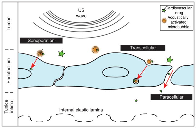
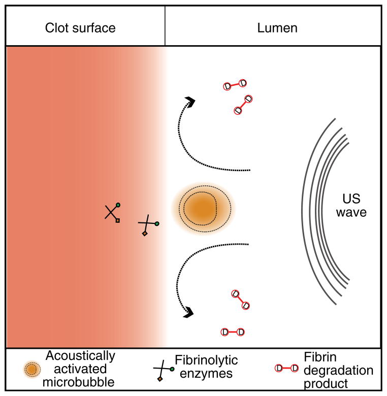
Introduction: Ultrasound (US) has been developed as both a valuable diagnostic tool and a potent promoter of beneficial tissue bioeffects for the treatment of cardiovascular disease. These effects can be mediated by mechanical oscillations of circulating microbubbles, or US contrast agents, which may also encapsulate and shield a therapeutic agent in the bloodstream. Oscillating microbubbles can create stresses directly on nearby tissue or induce fluid effects that effect drug penetration into vascular tissue, lyse thrombi or direct drugs to optimal locations for delivery.

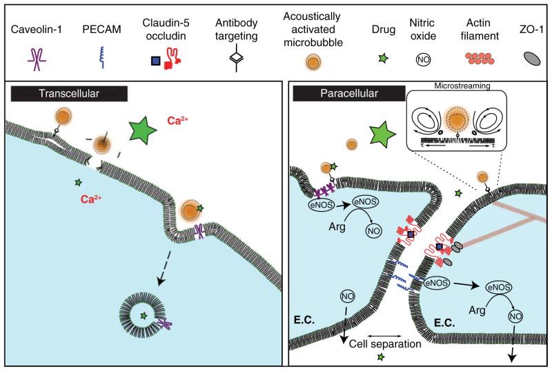
Areas covered: The present review summarizes investigations that have provided evidence for US-mediated drug delivery as a potent method to deliver therapeutics to diseased tissue for cardiovascular treatment. In particular, the focus will be on investigations of specific aspects relating to US-mediated drug delivery, such as delivery vehicles, drug transport routes, biochemical mechanisms and molecular targeting strategies.

Expert opinion: These investigations have spurred continued research into alternative therapeutic applications, such as bioactive gas delivery and new US technologies. Successful implementation of US-mediated drug delivery has the potential to change the way many drugs are administered systemically, resulting in more effective and economical therapeutics, and less-invasive treatments.


Conflict of interest statement

Declaration of interest

Grant funding from NIH R01 NS047603; NIH R01 HL059586; NIH R01 HL74002; NIH F32 HL104916 supported the coauthors during the preparation of this manuscript.

Figures

Figure 1
Figure 1. An overview of three penetration routes stimulated for ultrasound-mediated drug delivery
Sonoporation refers to the localized, mechanical disruption of a plasma membrane, which allows drugs and ions to diffuse passively. Transcellular pathways, such as endocytosis, involve active transport of drug via cytosolic vesicles. The paracellular route occurs when endothelial cells spread apart, either due to desquamation or by tight junction breakdown from bubble-induced shear stress.
Figure 2
Figure 2. An overview of some proposed mechanisms for transcellular and paracellular ultrasound-mediated drug delivery
Transcellular: as a result of bubble-induced shear stress along the cell membrane, extracellular drugs can undergo caveolin-1-mediated endocytosis. Additionally, sonoporation can create ‘holes’ in the cell membrane facilitating influx of ions or drugs. Paracellular: shear stress from cavitation-induced microstreaming can cause caveolin-1 to detach from endothelial nitric oxide synthase (eNOS). Here, it converts arginine to nitric oxide, stimulating vasodilation and possible paracellular permeability. Alternatively, this shear stress deforms the actin cytoskeleton, which can cause conformational changes and breakdown of tight junction proteins (ZO-1, occludin).
Figure 3
Figure 3. Propidium iodide (PI; left column) and intracellular [Ca2+]i flux (right column) during microbubble oscillation near a cell membrane
Cell outlines, indicated by white lines, demarcate PI and Ca2+ delivery, when a bubble (circle) oscillates near its cell membrane. The initial state of the cell is seen in the first row, followed by subsequent images during ultrasound exposure. Reprinted from Fan et al. [133], with permission from Elsevier (2012).
Figure 4
Figure 4. Images of calcein fluorescence indicating penetration of liposomes targeting to smooth muscle actin, in the absence (left) and presence (right) of ultrasound
Reprinted from Laing et al. [112], with permission from Informa Healthcare (2012).
Figure 5
Figure 5. Cavitational mechanisms in sonothrombolysis
As a result of local fluid dynamics around a cavitating microbubbles, the fibrinolytic enzymes (recombinant tissue-plasminogen activator (rt-PA) and plasminogen) penetrate deeper into the fibrin matrix. Additionally, fluid microstreaming removes fibrin degradation products from the surface of the clot, expediting the fibrinolytic process.

References

    1. Komarova Y, Malik A. Regulation of endothelial permeability via paracellular and transcellular transport pathways. Annu Rev Physiol. 2010;72(1):463–93. - PubMed
    1. Zaragoza C, Márquez S, Saura M. Endothelial mechanosensors of shear stress as regulators of atherogenesis. Curr Opin Lipidol. 2012;23(5):446–52. - PubMed
    1. Binsalamah Z, Paul A, Prakash S, Shum-Tim D. Nanomedicine in cardiovascular therapy: recent advancements. Expert Rev Cardiovasc Ther. 2012;10(6):805–15. - PubMed
    1. Roger V, Go A, Lloyd-Jones D, et al. Heart disease and stroke statistics –2012 update: a report from the American Heart Association. Circulation. 2012;125(1):e2–e220. - PMC - PubMed
    1. Stancu C, Toma L, Sima A. Dual role of lipoproteins in endothelial cell dysfunction in atherosclerosis. Cell Tissue Res. 2012;349(2):433–46. - PubMed

Publication types

MeSH terms

Substances

LinkOut - more resources