. 2013 Dec;8(12):1943-54.
doi: 10.2217/nnm.12.200. Epub 2013 Feb 28.

Tetraiodothyroacetic acid-conjugated PLGA nanoparticles: a nanomedicine approach to treat drug-resistant breast cancer

Affiliations

Tetraiodothyroacetic acid-conjugated PLGA nanoparticles: a nanomedicine approach to treat drug-resistant breast cancer

Dhruba J Bharali et al. Nanomedicine (Lond). 2013 Dec.

Retraction in

Abstract

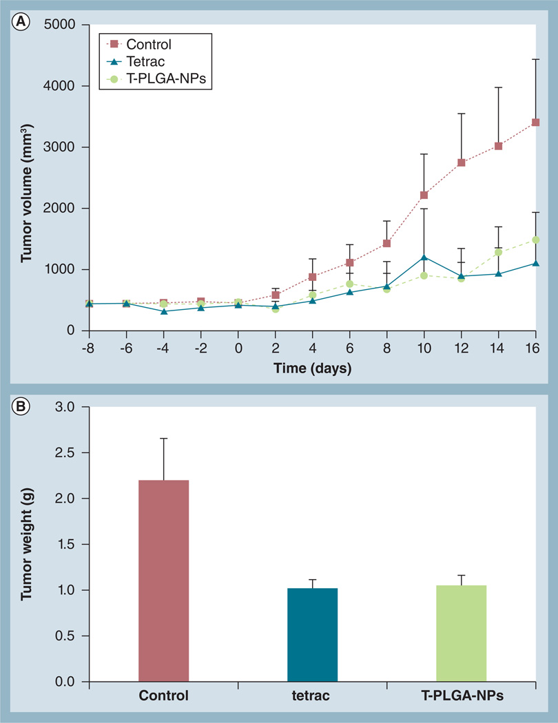
Aim: The aim was to evaluate tetraiodothyroacetic acid (tetrac), a thyroid hormone analog of L-thyroxin, conjugated to poly(lactic-co-glycolic acid) nanoparticles (T-PLGA-NPs) both in vitro and in vivo for the treatment of drug-resistant breast cancer.

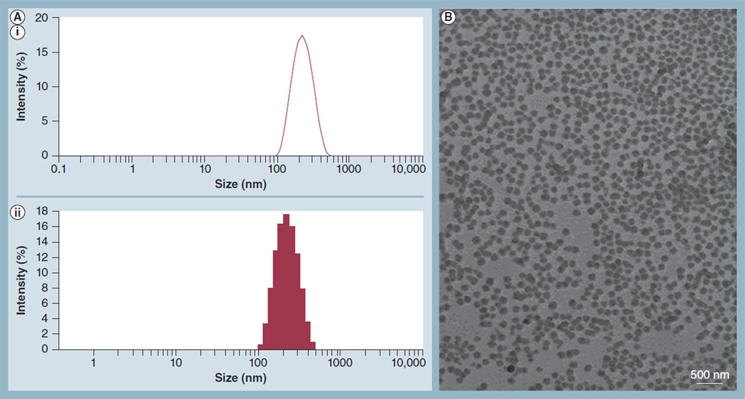
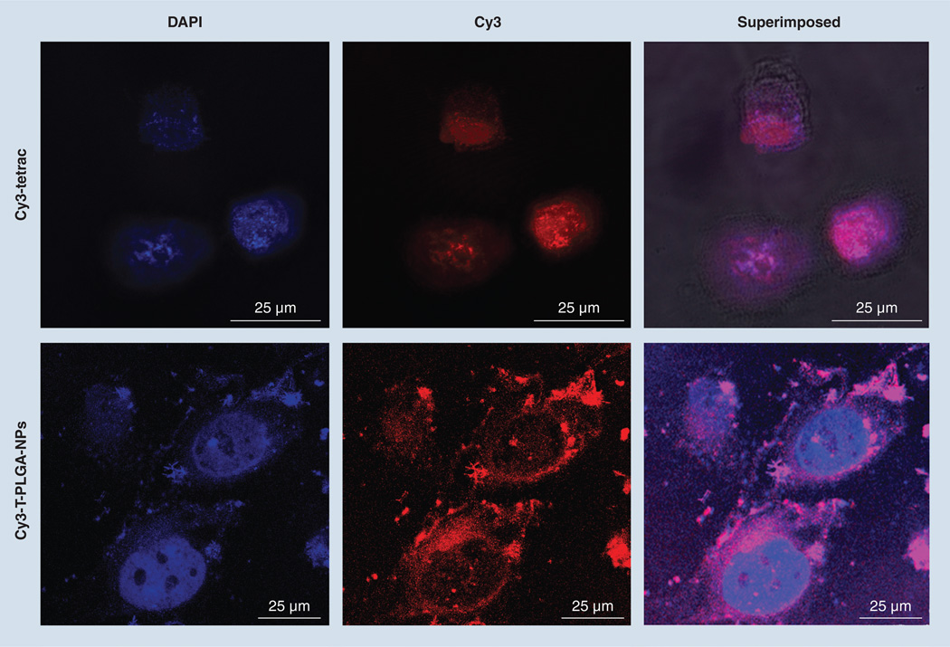
Materials & methods: The uptake of tetrac and T-PLGA-NPs in doxorubicin-resistant MCF7 (MCF7-Dx) cells was evaluated using confocal microscopy. Cell proliferation assays and a chick chorioallantoic membrane model of FGF2-induced angiogenesis were used to evaluate the anticancer effects of T-PLGA-NPs. In vivo efficacy was examined in a MCF7-Dx orthotopic tumor BALBc nude mouse model.

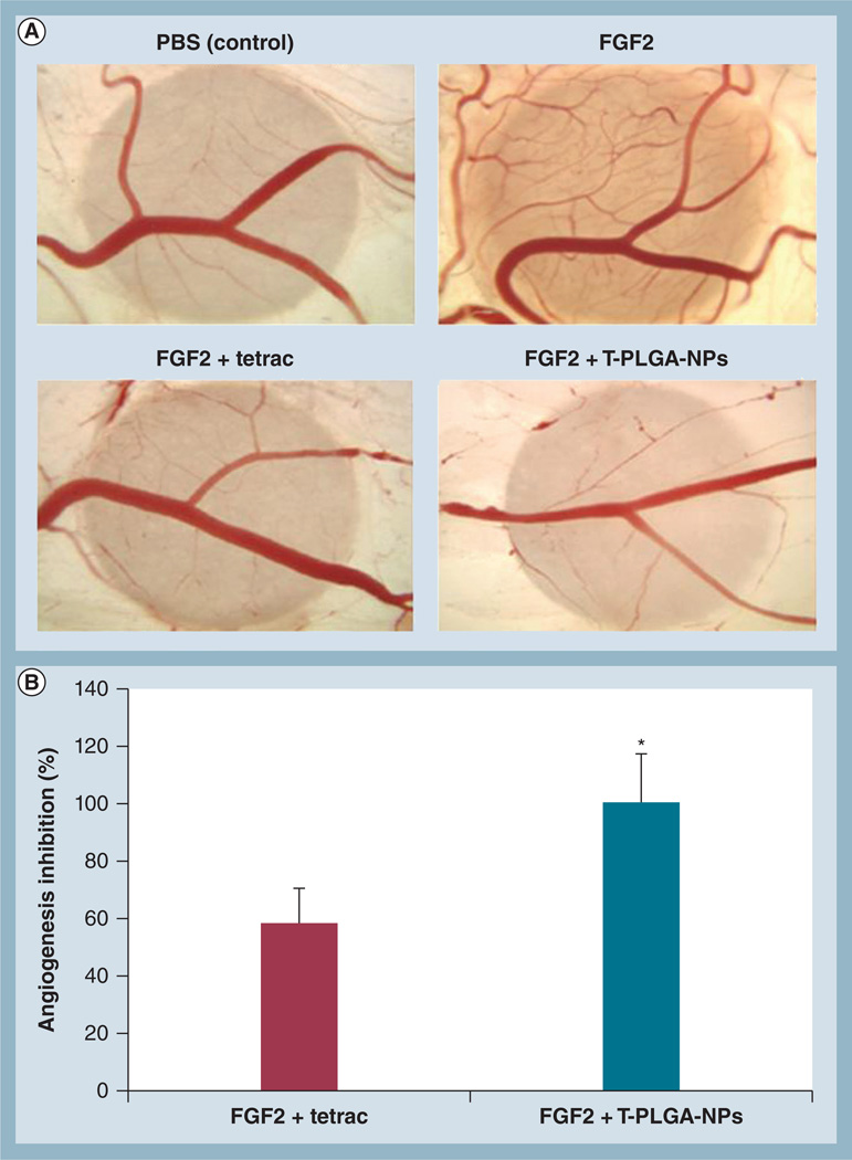
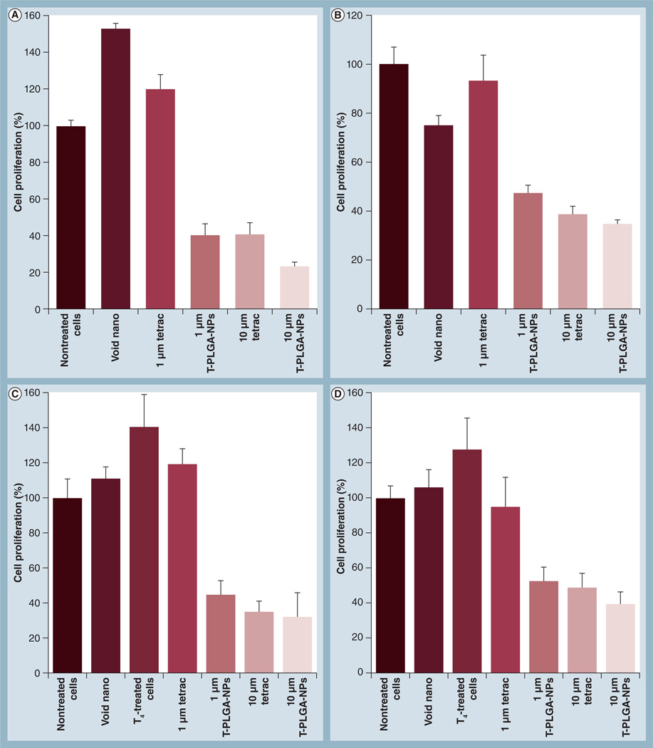
Results: T-PLGA-NPs were restricted from entering into the cell nucleus, and T-PLGA-NPs inhibited angiogenesis by 100% compared with 60% by free tetrac. T-PLGA-NPs enhanced inhibition of tumor-cell proliferation at a low-dose equivalent of free tetrac. In vivo treatment with either tetrac or T-PLGA-NPs resulted in a three- to five-fold inhibition of tumor weight.

Conclusion: T-PLGA-NPs have high potential as anticancer agents, with possible applications in the treatment of drug-resistant cancer.


Figures

Figure 1
Figure 1. Measurement of nanoparticle size and zeta-potential by dynamic light scattering and transmission electron microscopy
(A) A representative histogram from dynamic light scattering data of the size (diameter) distribution of tetraiodothyroacetic acid-conjugated poly(lactic-co-glycolic acid) nanoparticles. The average size of the tetraiodothyroacetic acid-conjugated poly(lactic-co-glycolic acid) nanoparticles was approximately 200 nm; (AI) size distribution by intensity; and (AII) intensity statistics. (B) Transmission electron microscopy image showing the morphology of tetraiodothyroacetic acid-conjugated poly(lactic-co-glycolic acid) nanoparticles.
Figure 2
Figure 2. Confocal microscopy images showing the uptake of Cy3-tetraiodothyroacetic acid and Cy3-tetraiodothyroacetic acid-conjugated poly(lactic-co-glycolic acid) nanoparticles by doxorubicin-resistant MCF7 breast cancer cells
Magnifcation: 63×; Optical zoom: 5×. Cells were counter-stained with DAPI to visualize the nuclei: blue for DAPI and red for Cy3-tetrac or Cy3-T-PLGA–NPs. DAPI: 4’,6-diamidino-2-phenylindole fluorescent stain; tetrac: Tetraiodothyroacetic acid; T-PLGA-NPs: Tetraiodothyroacetic acid conjugated to poly(lactic-co-glycolic acid) nanoparticles.
Figure 3
Figure 3. The effects of tetraiodothyroacetic acid-conjugated poly(lactic-co-glycolic acid) nanoparticles on FGF2-induced angiogenesis measured by the chick chorioallantoic membrane assay
(A) Photomicrographs of representative chick chorioallantoic membranes treated with free tetrac or T-PLGA-NPs. Chick chorioallantoic membranes were also treated with FGF2 or PBS as a positive and a negative control, respectively. (B) Quantification of angiogenesis inhibition by tetrac versus T-PLGA-NPs. Data represent the number of branch points and were normalized to the negative control (PBS). Data are expressed as percentage inhibition of angiogenesis induced by FGF2 alone (mean ± standard error of the mean). *p ≤ 0.01. PBS: Phosphate-buffered saline; tetrac: Tetraiodothyroacetic acid; T-PLGA-NPs: Tetraiodothyroacetic acid conjugated to poly(lactic-co-glycolic acid) nanoparticles.
Figure 4
Figure 4. Cell proliferation assays of tetraiodothyroacetic acid-conjugated poly(lactic-co-glycolic acid) nanoparticles showing the inhibition of cell proliferation
(A) MCF7 breast cancer cells. (B) Doxorubicin-resistant MCF7 (MCF7-Dx) breast cancer cells. (C) MCF7 breast cancer cells, incubated with 1 µM T4(D) MCF7-Dx breast cancer cells, incubated with 1 µM T4. A low dose (1 µM) of free tetrac has no effect on cell proliferation in either MCF7 or MCF7-Dx cells, whereas even a low-dose equivalent of tetrac conjugated to nanoparticles can inhibit cell proliferation by 60%. A high dose (10 µM) of free tetrac has a similar effect on inhibition to that of a high dose of tetrac conjugated to nanoparticles. T4: l-thyroxin; tetrac: Tetraiodothyroacetic acid; T-PLGA-NPs: Tetraiodothyroacetic acid-conjugated to poly(lactic-co-glycolic acid) nanoparticles; Void nano: Nanoparticles only, without any tetrac.
Figure 5
Figure 5. In vivo suppression by tetraiodothyroacetic acid versus tetraiodothyroacetic acid-conjugated poly(lactic-co-glycolic acid) nanoparticles in drug-resistant breast cancer orthotopic tumor mouse model
Treatment with tetrac and T-PLGA-NPs (1 mg/kg bodyweight) was started on day 8, after orthotopically implanting doxorubicin-resistant MCF7 cells in female BALBc nude mice, and continued daily. (A) Effect of tetrac and T-PLGA-NP treatment on doxorubicin-resistant MCF7 tumor volume over time, up to 16 days. Data represent mean tumor volume (mm3) ± standard deviation; n = 8 per group. (B) Effect on tumor weight at day 16 after the treatments (end of study). tetrac: Tetraiodothyroacetic acid; T-PLGA-NPs: Tetraiodothyroacetic acid conjugated to poly(lactic-co-glycolic acid) nanoparticles.

Comment in

  • Findings of Research Misconduct.
    [No authors listed] [No authors listed] Fed Regist. 2024 Jun 4;89(108):47968-47969. Fed Regist. 2024. PMID: 38854442 Free PMC article. No abstract available.

References

    1. Bharali DJ, Mousa SA. Emerging nanomedicines for early cancer detection, improved treatment: current perspective future promise. Pharmacol. Ther. 2010;128(2):324–335. - PubMed
    1. Brannon-Peppas L, Blanchette JO. Nanoparticle targeted systems for cancer therapy. Adv. Drug Deliv. Rev. 2004;56(11):1649–1659. - PubMed
    1. Nagahara LA, Lee JS, Molnar LK, et al. Strategic workshops on cancer nanotechnology. Cancer Res. 2010;70(11):4265–4268. - PMC - PubMed
    1. Jain RK, Stylianopoulos T. Delivering nanomedicine to solid tumors. Nat. Rev. Clin. Oncol. 2010;7(11):653–664. - PMC - PubMed
    1. Kim K, Kim JH, Park H, et al. Tumor-homing multifunctional nanoparticles for cancer theragnosis: simultaneous diagnosis drug delivery therapeutic monitoring. J. Control. Release. 2010;146(2):219–227. - PubMed

Publication types

MeSH terms