. 2013;19(34):6218-25.
doi: 10.2174/1381612811319340013.

Bioactivity of turmeric-derived curcuminoids and related metabolites in breast cancer

Affiliations

Bioactivity of turmeric-derived curcuminoids and related metabolites in breast cancer

Laura E Wright et al. Curr Pharm Des. 2013.

Abstract

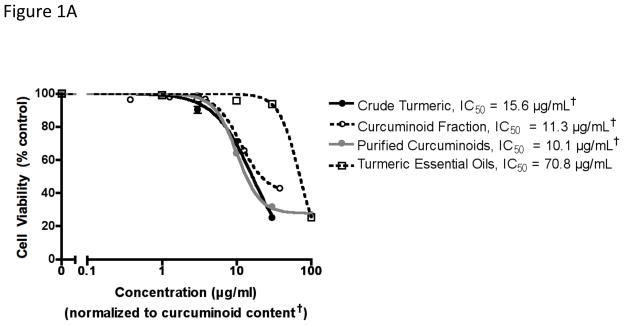
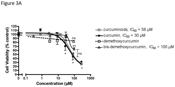
While the chemotherapeutic effect of curcumin, one of three major curcuminoids derived from turmeric, has been reported, largely unexplored are the effects of complex turmeric extracts more analogous to traditional medicinal preparations, as well as the relative importance of the three curcuminoids and their metabolites as anti-cancer agents. These studies document the pharmacodynamic effects of chemically-complex turmeric extracts relative to curcuminoids on human breast cancer cell growth and tumor cell secretion of parathyroid hormone-related protein (PTHrP), an important driver of cancer bone metastasis. Finally, relative effects of structurallyrelated metabolites of curcuminoids were assessed on the same endpoints. We report that 3 curcuminoid-containing turmeric extracts differing with respect to the inclusion of additional naturally occurring chemicals (essential oils and/or polar compounds) were equipotent in inhibiting human breast cancer MDA-MB-231 cell growth (IC50=10-16µg/mL) and secretion of osteolytic PTHrP (IC50=2-3µg/mL) when concentrations were normalized to curcuminoid content. Moreover, these effects were curcuminoid-specific, as botanically-related gingerol containing extracts had no effect. While curcumin and bis-demethoxycurcumin were equipotent to each other and to the naturally occurring curcuminoid mixture (IC50=58µM), demethoxycurcumin did not have any effect on cell growth. However, each of the individual curcuminoids inhibited PTHrP secretion (IC50=22-31µM) to the same degree as the curcuminoid mixture (IC50=16µM). Degradative curcuminoid metabolites (vanillin and ferulic acid) did not inhibit cell growth or PTHrP, while reduced metabolites (tetrahydrocurcuminoids) had inhibitory effects on cell growth and PTHrP secretion but only at concentrations ≥10-fold higher than the curcuminoids. These studies emphasize the structural and biological importance of curcuminoids in the anti-breast cancer effects of turmeric and contradict recent assertions that certain of the curcuminoid metabolites studied here mediate these anti-cancer effects.


Figures

Figure 1
Figure 1
Effect of chemically complex turmeric extracts and fractions on human breast cancer MDA-MB-231 cell viability, cytotoxicity and secretion of osteolytic PTHrP. As indicated in the methods, MDA-MB-231 cells (n = 4–8/group) were treated with botanicals for 4 hours prior to stimulation with TGFβ for 24 hours, after which time (A) cell viability was assessed by MTT assay and (B) PTHrP secretion was determined by radioimmunometric assay of conditioned media. Concentrations of all curcuminoid-containing extracts were normalized to curcuminoid content (†). Half maximal inhibitory concentrations (IC50) were obtained using a four-parameter logistic equation with statistically significant differences between IC50 values determined by F test with p < 0.05. (C) Effects of purified curcuminoids (5 μg/ml) and turmeric essential oils (5 μg/ml) alone or in combination on PTHrP secretion (n = 4/group), and (D) effects of purified curcuminoids on cell viability (MTT assay) vs. cytotoxicity (LDH content of media) (n = 4/group) were compared with statistically significant effects determined by ANOVA with post-hoc testing. *p < 0.05 vs. control. ***p < 0.001 vs control.
Figure 1
Figure 1
Effect of chemically complex turmeric extracts and fractions on human breast cancer MDA-MB-231 cell viability, cytotoxicity and secretion of osteolytic PTHrP. As indicated in the methods, MDA-MB-231 cells (n = 4–8/group) were treated with botanicals for 4 hours prior to stimulation with TGFβ for 24 hours, after which time (A) cell viability was assessed by MTT assay and (B) PTHrP secretion was determined by radioimmunometric assay of conditioned media. Concentrations of all curcuminoid-containing extracts were normalized to curcuminoid content (†). Half maximal inhibitory concentrations (IC50) were obtained using a four-parameter logistic equation with statistically significant differences between IC50 values determined by F test with p < 0.05. (C) Effects of purified curcuminoids (5 μg/ml) and turmeric essential oils (5 μg/ml) alone or in combination on PTHrP secretion (n = 4/group), and (D) effects of purified curcuminoids on cell viability (MTT assay) vs. cytotoxicity (LDH content of media) (n = 4/group) were compared with statistically significant effects determined by ANOVA with post-hoc testing. *p < 0.05 vs. control. ***p < 0.001 vs control.
Figure 2
Figure 2
Effect of chemically complex ginger extracts and fractions on human breast cancer MDA-MB-231 cell viability and secretion of osteolytic PTHrP. As indicated in the methods, MDA-MB-231 cells (n = 4–8/group) were treated with botanicals for 4 hours prior to stimulation with TGFβ for 24 hours, after which time (A) cell viability was assessed by MTT assay and (B) PTHrP secretion was determined by radioimmunometric assay of conditioned media. Concentrations of all gingerol-containing extracts were normalized to gingerol content (†). Half maximal inhibitory concentrations (IC50) were obtained using a four-parameter logistic equation with statistically significant differences between IC50 values determined by F test with p < 0.05.
Figure 2
Figure 2
Effect of chemically complex ginger extracts and fractions on human breast cancer MDA-MB-231 cell viability and secretion of osteolytic PTHrP. As indicated in the methods, MDA-MB-231 cells (n = 4–8/group) were treated with botanicals for 4 hours prior to stimulation with TGFβ for 24 hours, after which time (A) cell viability was assessed by MTT assay and (B) PTHrP secretion was determined by radioimmunometric assay of conditioned media. Concentrations of all gingerol-containing extracts were normalized to gingerol content (†). Half maximal inhibitory concentrations (IC50) were obtained using a four-parameter logistic equation with statistically significant differences between IC50 values determined by F test with p < 0.05.
Figure 3
Figure 3
Comparative effects of curcuminoids, curcumin, demethoxycurcumin and bis-demethoxycurcumin on human breast cancer MDA-MB-231 cell viability and secretion of osteolytic PTHrP. MDA-MB-231 cells (n = 8–20/group) were treated with botanicals for 4 hours prior to stimulation with TGFβ for 24 hours, after which time (A) cell viability was assessed by MTT assay and (B) PTHrP secretion was determined by radioimmunometric assay of conditioned media. Half maximal inhibitory concentrations (IC50) were obtained using a four-parameter logistic equation with statistically significant differences between IC50 values determined by F test with p < 0.05. Statistically significant effects of each botanical (vs. controls or each other) were also determined by ANOVA with post hoc testing. * p < 0.001 (vs. control) for curcumin or curcuminoids. ** p < 0.001 (vs. control) for bis-demethoxycurcumin.
Figure 3
Figure 3
Comparative effects of curcuminoids, curcumin, demethoxycurcumin and bis-demethoxycurcumin on human breast cancer MDA-MB-231 cell viability and secretion of osteolytic PTHrP. MDA-MB-231 cells (n = 8–20/group) were treated with botanicals for 4 hours prior to stimulation with TGFβ for 24 hours, after which time (A) cell viability was assessed by MTT assay and (B) PTHrP secretion was determined by radioimmunometric assay of conditioned media. Half maximal inhibitory concentrations (IC50) were obtained using a four-parameter logistic equation with statistically significant differences between IC50 values determined by F test with p < 0.05. Statistically significant effects of each botanical (vs. controls or each other) were also determined by ANOVA with post hoc testing. * p < 0.001 (vs. control) for curcumin or curcuminoids. ** p < 0.001 (vs. control) for bis-demethoxycurcumin.
Figure 4
Figure 4
Chemical structures of curcumin and related compounds tested.
Figure 5
Figure 5
Comparative effects of curcuminoids, tetrahydrocurcuminoids, ferulic acid, vanillin, and 6-gingerol on human breast cancer MDA-MB-231 cell viability and secretion of osteolytic PTHrP. MDA-MB-231 cells (n = 8–16 per group) were treated with botanicals for 4 hours prior to stimulation with TGFβ for 24 hours, after which time (A) cell viability was assessed by MTT assay and (B) PTHrP secretion was determined by radioimmunometric assay of conditioned media. Half maximal inhibitory concentrations (IC50) were obtained using a four-parameter logistic equation with statistically significant differences between IC50 values determined by F test with p < 0.05. For each compound, statistically significant effects (vs. control) were determined by ANOVA with post-hoc testing. *p < 0.05. ** p < 0.01. ***p < 0.001.
Figure 5
Figure 5
Comparative effects of curcuminoids, tetrahydrocurcuminoids, ferulic acid, vanillin, and 6-gingerol on human breast cancer MDA-MB-231 cell viability and secretion of osteolytic PTHrP. MDA-MB-231 cells (n = 8–16 per group) were treated with botanicals for 4 hours prior to stimulation with TGFβ for 24 hours, after which time (A) cell viability was assessed by MTT assay and (B) PTHrP secretion was determined by radioimmunometric assay of conditioned media. Half maximal inhibitory concentrations (IC50) were obtained using a four-parameter logistic equation with statistically significant differences between IC50 values determined by F test with p < 0.05. For each compound, statistically significant effects (vs. control) were determined by ANOVA with post-hoc testing. *p < 0.05. ** p < 0.01. ***p < 0.001.

Similar articles

Cited by

References

    1. Centers for Disease Control and Prevention. 1999–2008 Incidence and Mortality Web-Based Report. Atlanta GA: Department of Health and Human Services, Center for Disease Control and Prevention, and National Cancer Institute; [Accessed 26 Oct 2012]. homepage on the internet. Available from: http://www.cdc.gov/uscs.
    1. World Health Organization, International Agency for Research on Cancer. GLOBOCAN. Lyon, France: 2008. [Accessed 26 Oct 2012]. homepage on the internet. Available from: http://globocan.iarc.fr.
    1. Ji JL, Huang XF, Zhu HL. Curcumin and its formulations: potential anti-cancer agents. Anticancer Agents Med Chem. 2012;12:210–8. - PubMed
    1. Mundy GR. Metastasis to bone: causes, consequences and therapeutic opportunities. Nat Rev Cancer. 2002;8:584–93. - PubMed
    1. Wright LE, Frye JB, Lukefahr AL, et al. Curcuminoids block TGF-β signaling in human breast cancer cells and limit osteolysis in a murine model of breast cancer bone metastasis. J Nat Prod. 2012 (in press) - PMC - PubMed

Publication types

MeSH terms