

Structural studies of the interaction of Crataeva tapia bark protein with heparin and other glycosaminoglycans
- PMID: 23448527
- PMCID: PMC3855636
- DOI: 10.1021/bi400077b
Structural studies of the interaction of Crataeva tapia bark protein with heparin and other glycosaminoglycans
Abstract
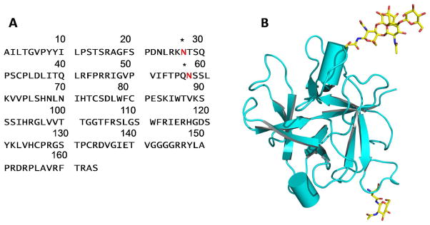
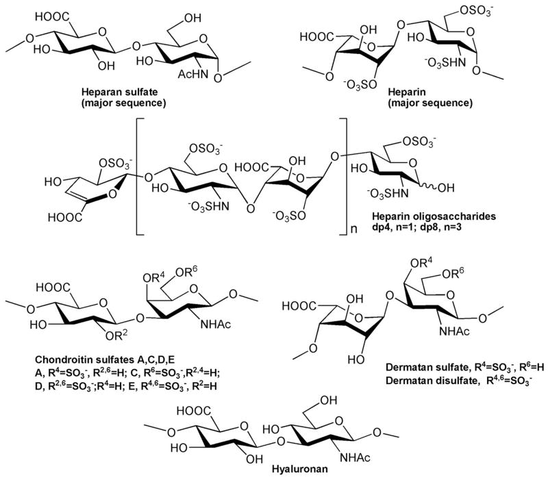
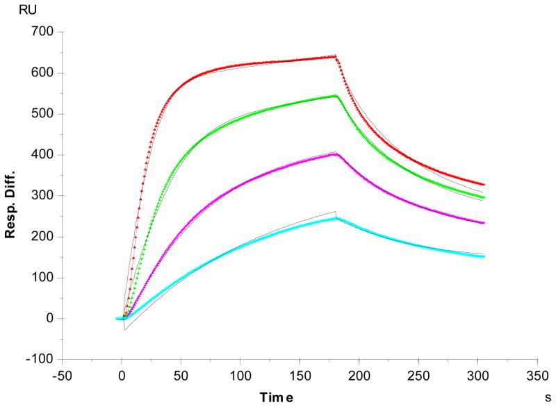
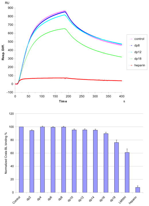
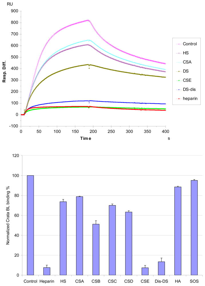
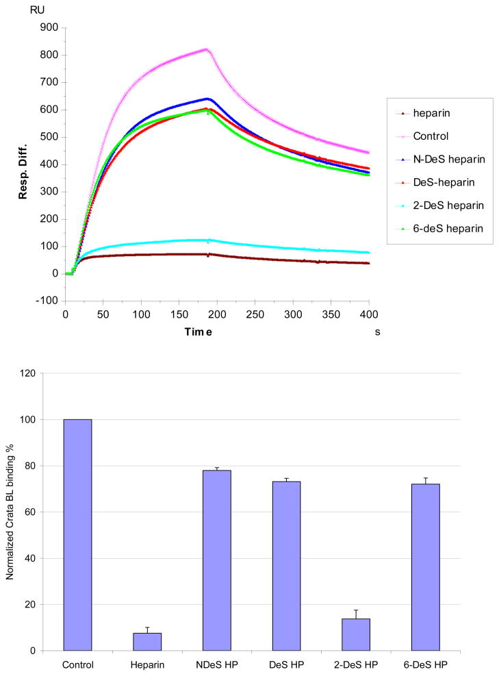
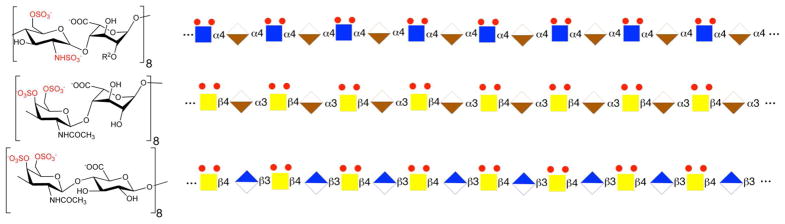
CrataBL, a protein isolated from Crataeva tapia bark, which is both a serine protease inhibitor and a lectin, has been previously shown to exhibit a number of interesting biological properties, including anti-inflammatory, analgesic, antitumor, and insecticidal activities. Using a glycan array, we have now shown that only sulfated carbohydrates are effectively bound by CrataBL. Because this protein was recently shown to delay clot formation by impairing the intrinsic pathway of the coagulation cascade, we considered that its natural ligand might be heparin. Heparin is a glycosaminoglycan (GAG) that interacts with a number of proteins, including thrombin and antithrombin III, which have a critical, essential pharmacological role in regulating blood coagulation. We have thus employed surface plasmon resonance to improve our understanding of the binding interaction between the heparin polysaccharide and CrataBL. Kinetic analysis shows that CrataBL displays strong heparin binding affinity (KD = 49 nM). Competition studies using different size heparin-derived oligosaccharides showed that the binding of CrataBL to heparin is chain length-dependent. Full chain heparin with 40 saccharides or large oligosaccharides, having 16-18 saccharide residues, show strong binding affinity for CrataBL. Heparin-derived disaccharides through tetradecasaccharides show considerably lower binding affinity. Other highly sulfated GAGs, including chondroitin sulfate E and dermatan 4,6-disulfate, showed CrataBL binding affinity comparable to that of heparin. Less highly sulfated GAGs, heparan sulfate, chondroitin sulfate A and C, and dermatan sulfate displayed modest binding affinity as did chondroitin sulfate D. Studies using chemically modified heparin show that N-sulfo and 6-O-sulfo groups on heparin are essential for CrataBL-heparin interaction.
Conflict of interest statement
The authors declare no competing financial interest.
Figures
Similar articles
-
Characterization of the interaction between Robo1 and heparin and other glycosaminoglycans.Biochimie. 2013 Dec;95(12):2345-53. doi: 10.1016/j.biochi.2013.08.018. Epub 2013 Aug 28. Biochimie. 2013. PMID: 23994753 Free PMC article.
-
SPR Biosensor Probing the Interactions between TIMP-3 and Heparin/GAGs.Biosensors (Basel). 2015 Jul 23;5(3):500-12. doi: 10.3390/bios5030500. Biosensors (Basel). 2015. PMID: 26213979 Free PMC article.
-
Crataeva tapia bark lectin (CrataBL) is a chemoattractant for endothelial cells that targets heparan sulfate and promotes in vitro angiogenesis.Biochimie. 2019 Nov;166:173-183. doi: 10.1016/j.biochi.2019.04.011. Epub 2019 Apr 12. Biochimie. 2019. PMID: 30981871
-
The interaction of glycosaminoglycans with heparin cofactor II.Ann N Y Acad Sci. 1994 Apr 18;714:21-31. doi: 10.1111/j.1749-6632.1994.tb12027.x. Ann N Y Acad Sci. 1994. PMID: 8017769 Review.
-
Specificity of glycosaminoglycan-protein interactions.Curr Opin Struct Biol. 2018 Jun;50:101-108. doi: 10.1016/j.sbi.2017.12.011. Epub 2018 Feb 9. Curr Opin Struct Biol. 2018. PMID: 29455055 Review.
Cited by
-
Cutaneous Melanoma: An Overview of Physiological and Therapeutic Aspects and Biotechnological Use of Serine Protease Inhibitors.Molecules. 2024 Aug 16;29(16):3891. doi: 10.3390/molecules29163891. Molecules. 2024. PMID: 39202970 Free PMC article. Review.
-
Crystal Structure of Crataeva tapia Bark Protein (CrataBL) and Its Effect in Human Prostate Cancer Cell Lines.PLoS One. 2013 Jun 18;8(6):e64426. doi: 10.1371/journal.pone.0064426. Print 2013. PLoS One. 2013. PMID: 23823708 Free PMC article.
-
Sources, Extraction and Biomedical Properties of Polysaccharides.Foods. 2019 Aug 1;8(8):304. doi: 10.3390/foods8080304. Foods. 2019. PMID: 31374889 Free PMC article. Review.
-
A Bifunctional Molecule with Lectin and Protease Inhibitor Activities Isolated from Crataeva tapia Bark Significantly Affects Cocultures of Mesenchymal Stem Cells and Glioblastoma Cells.Molecules. 2019 Jun 4;24(11):2109. doi: 10.3390/molecules24112109. Molecules. 2019. PMID: 31167364 Free PMC article.
-
Impairment of SK-MEL-28 Development-A Human Melanoma Cell Line-By the Crataeva tapia Bark Lectin and Its Sequence-Derived Peptides.Int J Mol Sci. 2023 Jun 25;24(13):10617. doi: 10.3390/ijms241310617. Int J Mol Sci. 2023. PMID: 37445794 Free PMC article.
References
-
- Varki A, Cummings RD, Esko JD, et al., editors. Essentials of Glycobiology. 2. Cold Spring Harbor (NY): Cold Spring Harbor Laboratory Press; 2009. - PubMed
-
- Correia MTS, Coelho LCBB, Paiva PMG. Lectins, carbohydrate recognition molecules: are they toxic? In: Siddique YH, editor. Recent Trends in Toxicology. Vol. 37. Transworld Research Network; Kerala: 2008. pp. 47–59.
-
- Li YR, Liu QH, Wang HX, Ng TB. A novel lectin with potent antitumor, mitogenic and HIV-1 reverse transcriptase inhibitory activities from the edible mushroom Pleurotus citrinopileatus. Biochim Biophys Acta: Gen Subjects. 2008;1780:51–57. - PubMed
-
- Capila I, Linhardt RJ. Heparin-protein interactions. AngewandteChemie-Intern Ed. 2002;41:391–412. - PubMed
-
- Hacker U, Nybakken K, Perrimon N. Heparansulphate proteoglycans: the sweet side of development. Nat Rev Mol Cell Bio. 2005;6:530–541. - PubMed
Publication types
MeSH terms
Substances
Grants and funding
LinkOut - more resources
Full Text Sources
Other Literature Sources
Medical
