. 2013 Mar 1:13:96.
doi: 10.1186/1471-2407-13-96.

Bcl-2 associated athanogene 5 (Bag5) is overexpressed in prostate cancer and inhibits ER-stress induced apoptosis

Affiliations

Bcl-2 associated athanogene 5 (Bag5) is overexpressed in prostate cancer and inhibits ER-stress induced apoptosis

Anja Bruchmann et al. BMC Cancer. .

Abstract

Background: The Bag (Bcl-2 associated athanogene) family of proteins consists of 6 members sharing a common, single-copied Bag domain through which they interact with the molecular chaperone Hsp70. Bag5 represents an exception in the Bag family since it consists of 5 Bag domains covering the whole protein. Bag proteins like Bag1 and Bag3 have been implicated in tumor growth and survival but it is not known whether Bag5 also exhibits this function.

Methods: Bag5 mRNA and protein expression levels were investigated in prostate cancer patient samples using real-time PCR and immunoblot analyses. In addition immunohistological studies were carried out to determine the expression of Bag5 in tissue arrays. Analysis of Bag5 gene expression was carried out using one-way ANOVA and Bonferroni's Multiple Comparison test. The mean values of the Bag5 stained cells in the tissue array was analyzed by Mann-Whitney test. Functional studies of the role of Bag5 in prostate cancer cell lines was performed using overexpression and RNA interference analyses.

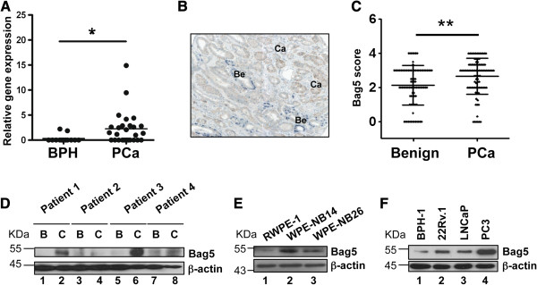
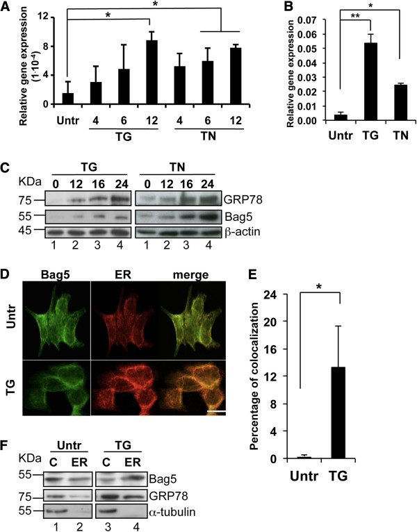
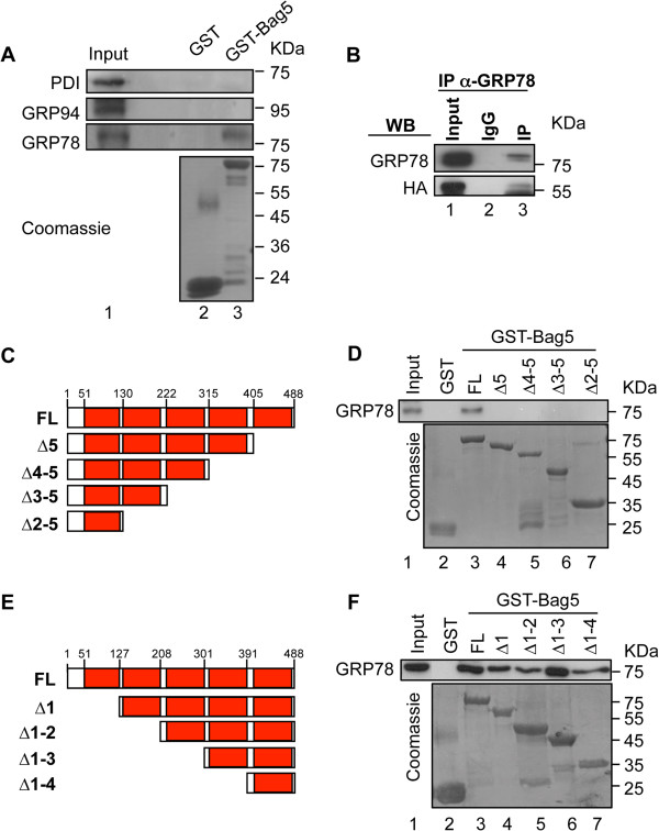
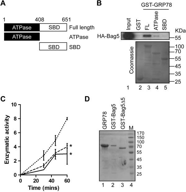
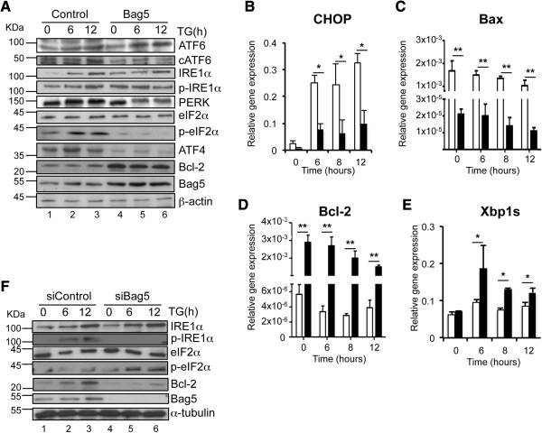
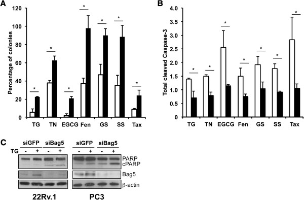
Results: Our results show that Bag5 is overexpressed in malignant prostate tissue compared to benign samples. In addition we could show that Bag5 levels are increased following endoplasmic reticulum (ER)-stress induction, and Bag5 relocates from the cytoplasm to the ER during this process. We also demonstrate that Bag5 interacts with the ER-resident chaperone GRP78/BiP and enhances its ATPase activity. Bag5 overexpression in 22Rv.1 prostate cancer cells inhibited ER-stress induced apoptosis in the unfolded protein response by suppressing PERK-eIF2-ATF4 activity while enhancing the IRE1-Xbp1 axis of this pathway. Cells expressing high levels of Bag5 showed reduced sensitivity to apoptosis induced by different agents while Bag5 downregulation resulted in increased stress-induced cell death.

Conclusions: We have therefore shown that Bag5 is overexpressed in prostate cancer and plays a role in ER-stress induced apoptosis. Furthermore we have identified GRP78/BiP as a novel interaction partner of Bag5.


Figures

Figure 1
Figure 1
Bag5 is overexpressed in prostate cancer. Bag5 expression analysis in prostate cancer tissue and cell lines. A. Quantitative RT-PCR studies showing the relative expression of Bag5 in benign prostate hyperplasia (BPH, n = 15) and prostate cancer (PCa, n = 27). The expression values were normalized against the expression of β-actin. The horizontal line indicates arithmetic mean value. Non-parametric Kruskal-Wallis with Dunn’ post-test was used to analyze the statistical significance of the differences between the groups (* = p <0.05). B. Representative immunohistochemical staining used in the tissue array analysis. P63 (blue) and Bag5 (brown) staining are shown. Bening (Be) and cancer (Ca) tissue is indicated. C. Tissue microarray containing tissue cores of 91 cancer cases. The horizontal line indicates arithmetic mean value. (**p < 0.001). D. Western blot analysis of Bag5 protein expression in prostate cancer patients comparing cancer (C) and surrounding benign tissue (B). β-actin was used as equal loading control. E. Western blot analysis of Bag5 protein expression in an in vitro prostate tumor progression model using RWPE-1 (benign), WPE-NB14 (primary tumor) and WPE-NB26 (metastatic tumor) cells. β-actin was used as equal loading control. F. Western blot analysis of Bag5 protein expression in prostate cell lines. Equal loading control was checked with β-actin.
Figure 2
Figure 2
Bag5 is stress-induced and accumulates in the endoplasmic reticulum. Bag5 gene expression is induced upon stress. A. Real time PCR analysis of Bag5 gene in 22Rv.1 cells upon treatment with thapsigargin (TG) or tunicamycin (TN) for the indicated time points. Gene expression values were normalized to Rib36. Bar charts indicate the mean value of three independent experiments ± SD. (* p < 0.05) B. Real time PCR analysis of GRP78 gene in 22Rv.1 cells upon treatment with thapsigargin (TG) or tunicamycin (TN) for 12 h. Gene expression values were normalized to Rib36. Bar charts indicate the mean value of three independent experiments ± SD. (* p < 0.05; ** p < 0.01). C. Bag5 protein expression is induced upon stress. Western blot analysis of 22Rv.1 cell extracts after thapsigargin or tunicamycin treatment for the indicated time points. β-actin was detected as loading control. D. Bag5 accumulates into the ER upon stress. D. Bag5 associates to the ER upon stress. Confocal microscopic analysis of 22Rv.1 cells-paraformaldehyde-fixed and stained with an anti-Bag5 antibody (green channel) or the ER-tracker (red channel) after 12 hours thapsigargin treatment. All images (40X) were acquired with a Leica TCS SPE confocal microscope (Leica Microsystems). The scale bar indicates 25 μm. E. Quantification of the percentage of co-localization performed with the software Imaris CoLoc. Bar charts indicate the average of three fields of three independent experiments ± SD (* p < 0.01). F. ER extraction from 22Rv.1 cell lysates treated with TG for 8 h. Western blot analysis was performed with a Bag5 or a GRP78 specific antibody. Anti-α-tubulin was used for ER fractions quality control (C: cytoplasm; ER: endoplasmic reticulum).
Figure 3
Figure 3
Bag5 interacts with GRP78/BiP. A. Bag5 interacts with GRP78/BiP but not with GRP94 and PDI. GST-pull down assay performed incubating 10 μg of GST-Bag5 and 250 μg of 22Rv.1 cell lysate. Equal recombinant protein employed in the assay was checked by staining the membrane with coomassie. 5% of the lysate was loaded as input. B. Bag5 and GRP78 interact in vivo. Co-immunoprecipitation assay performed in HEK293 cells transfected with HA-Bag5. GRP78/BiP was immunoprecipitated with an anti-GRP78 specific antibody and IgG was used as negative control. Western blot analysis was performed with an anti-HA and anti-GRP78 antibody. C-F. The fifth Bag domain of Bag5 mediates the interaction with GRP78/BiP. C and E. Diagrammatic representation of Bag5 and the deletion mutants used for the GST-pull down assay. D and F. GST-pull down assay performed incubating 10 μg of GST-fused protein and 500 μg of 22Rv.1 cell lysate. An anti-GRP78 antibody was used for western blot analysis. The membrane was stained with coomassie for equal protein amount employed in the experiment.
Figure 4
Figure 4
Bag5 functions as co-chaperone of GRP78/BiP. A. Diagrammatic representation of the molecular chaperone GRP78/BiP and its domains. Numbers indicate the amino acid position. B. Bag5 binds the ATPase domain of GRP78. GST-pull down assay was performed incubating 10 μg of GST-fused protein with 500 μg of HEK-293 cells transfeced with HA-Bag5. A specific anti-HA antibody was used in western blot analysis to detect the binding. Equal recombinant protein employed in the assay was checked by coomassie staining. C. Bag5 enhances GRP78/BiP ATP hydrolysis. ATPase assay with purified GRP78 in absence or presence of Bag5 (dashed line) or Bag5∆ 5 (dotted line). Results are expressed as the average of three independent experiments ± SD (* p < 0.05) D. Coomassie staining of the proteins employed in the ATPase assay.
Figure 5
Figure 5
Bag5 overexpression modulates the unfolded protein response. A. 22Rv.1 stably overexpressing Bag5 showed increased IRE1/Xbp1 axis activation. Western blot analysis of 22Rv.1 cells stably transfected with pcDNA3.1-HA-Bag5 or the empty vector as control. After treatment with TG for the indicated time points, cells were lysed and subjected to western blot analysis using the indicated antibody. As equal loading control an anti-β-actin antibody was used. B - E. Real time-PCR analysis of 22Rv.1 cells stably expressing the empty vector control (open bars) or HA-Bag5 (filled bars) treated with TG for the indicated time points. Gene expression analysis was performed for CHOP (B), Bcl-2 (C), Bax (D) and the spliced form of Xbp1, Xbp1s (E). Values were normalized to Rib36. Bar charts indicate the mean of at least three independent experiments ± SD (*p <0.05; ** p < 0.01). F. Western blot analysis of 22Rv.1 cells transfected with siRNA targeting GFP or Bag5 and treated with TG for the indicated time points. Anti-α-tubulin was used as equal loading control.
Figure 6
Figure 6
Bag5 increases prostate cancer cell chemoresistance. Modulation of Bag5 levels influences prostate cancer cell response to chemotherapy. A and B. Colony forming assay (A) and caspase-3 cleavage analysis (B) of 22Rv.1 cells stably expressing the empty vector control (open bars) or HA-Bag5 (filled bars). Bar charts represent the average of three independent experiments ± SD. * p < 0.05. (TG: thapsigargin, 75 nM; TN: tunicamycin, 10 μg/ml; EGCG: (-)-epigallocatechingallate, 10 μM; Fen: fenretinide, 20 μM; GS: glucose starvation; SS: serum starvation; Tax: taxol, 10 nM). * p < 0.05; ** p < 0.01. C. Western blot analysis of 22Rv.1 and PC3 cells transfected with siRNA targeting Bag5 or GFP as control and treated with 300 nM TG for 24 hours. Anti-β-actin antibody was used as equal loading control.

References

    1. Doong H, Vrailas A, Kohn EC. What’s in the ‘BAG’?–A functional domain analysis of the BAG-family proteins. Cancer Lett. 2002;188(1–2):25–32. - PubMed
    1. Sondermann H, Scheufler C, Schneider C, Hohfeld J, Hartl FU, Moarefi I. Structure of a Bag/Hsc70 complex: convergent functional evolution of Hsp70 nucleotide exchange factors. Science. 2001;291(5508):1553–1557. doi: 10.1126/science.1057268. - DOI - PubMed
    1. Briknarova K, Takayama S, Brive L, Havert ML, Knee DA, Velasco J, Homma S, Cabezas E, Stuart J, Hoyt DW. Structural analysis of BAG1 cochaperone and its interactions with Hsc70 heat shock protein. Nat Struct Biol. 2001;8(4):349–352. doi: 10.1038/86236. - DOI - PubMed
    1. Brimmell M, Burns JS, Munson P, McDonald L, O’Hare MJ, Lakhani SR, Packham G. High level expression of differentially localized BAG-1 isoforms in some oestrogen receptor-positive human breast cancers. Br J cancer. 1999;81(6):1042–1051. doi: 10.1038/sj.bjc.6690805. - DOI - PMC - PubMed
    1. Briknarova K, Takayama S, Homma S, Baker K, Cabezas E, Hoyt DW, Li Z, Satterthwait AC, Ely KR. BAG4/SODD protein contains a short BAG domain. J Biol Chem. 2002;277(34):31172–31178. doi: 10.1074/jbc.M202792200. - DOI - PubMed

Publication types

MeSH terms