Case Reports
. 2013 Apr;22(2):76-80.
doi: 10.1097/MCD.0b013e32835fff39.

Early childhood presentation of Czech dysplasia

Affiliations
Case Reports

Early childhood presentation of Czech dysplasia

Lindsay C Burrage et al. Clin Dysmorphol. 2013 Apr.

Abstract

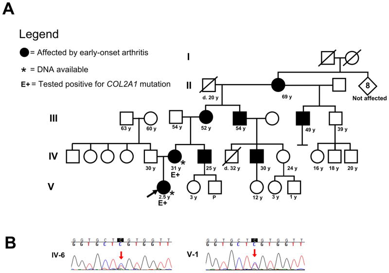
Czech dysplasia, metatarsal type is an autosomal dominant skeletal disorder that is characterized by early-onset, progressive arthritis, brachydactyly of the 3rd and 4th toes, and characteristic radiographic findings in patients of normal stature. Patients with Czech dysplasia typically present in late childhood or later. In the present report, whole exome sequencing identified a mutation in COL2A1 (c.823C>T, p.R275C) known to be associated with Czech dysplasia in a 3.5 year old female who had a family history of early-onset arthritis and who was asymptomatic except for prominent knees. The use of whole exome sequencing facilitated diagnosis of this rare disease (less than 15 families in the literature) in the presymptomatic period and thus enabled us to provide early anticipatory guidance and genetic counseling for the family.


Figures

Figure 1
Figure 1
A) Pedigree of the family described. B) Sanger sequencing results show that both the proband and her mother are heterozygous for the COL2A1 c.823C>T mutation C) Photographs of the patient, demonstrating enlarged knees and unremarkable feet, as was the case for her mother’s feet (D).
Figure 1
Figure 1
A) Pedigree of the family described. B) Sanger sequencing results show that both the proband and her mother are heterozygous for the COL2A1 c.823C>T mutation C) Photographs of the patient, demonstrating enlarged knees and unremarkable feet, as was the case for her mother’s feet (D).
Figure 2
Figure 2
Radiographs of the hands (A) and feet (B) of the proband show generalized shortening of the metacarpals and metatarsals. The proximal phalanges of the hands and feet are broad and short. The first metatarsal and the phalanges of the great toes are particularly wide. Frontal radiographs of lower extremities (C) demonstrate flattening of the distal femoral and the proximal and distal tibial epiphyses. Frontal and lateral spine radiographs in the proband (D) are normal. Radiographs of the hands (E) and feet (F) in the mother show no evidence of brachydactyly. There is evidence of generalized cartilage loss with joint space narrowing especially in the carpal and tarsal regions. In addition, there is widening of the metatarsal and phalanges of the great toe bilaterally. Frontal and lateral spine radiographs (G) in the mother show platyspondyly and Schmorl’s nodes. Frontal radiographs of the right shoulder (H) demonstrate an osteochondroma (arrow).
Figure 2
Figure 2
Radiographs of the hands (A) and feet (B) of the proband show generalized shortening of the metacarpals and metatarsals. The proximal phalanges of the hands and feet are broad and short. The first metatarsal and the phalanges of the great toes are particularly wide. Frontal radiographs of lower extremities (C) demonstrate flattening of the distal femoral and the proximal and distal tibial epiphyses. Frontal and lateral spine radiographs in the proband (D) are normal. Radiographs of the hands (E) and feet (F) in the mother show no evidence of brachydactyly. There is evidence of generalized cartilage loss with joint space narrowing especially in the carpal and tarsal regions. In addition, there is widening of the metatarsal and phalanges of the great toe bilaterally. Frontal and lateral spine radiographs (G) in the mother show platyspondyly and Schmorl’s nodes. Frontal radiographs of the right shoulder (H) demonstrate an osteochondroma (arrow).

References

    1. Bleasel JF, Bisagni-Faure A, Holderbaum D, Vacher-Lavenu MC, Haqqi TM, Moskowitz RW, Menkes CJ. Type II procollagen gene (COL2A1) mutation in exon 11 associated with spondyloepiphyseal dysplasia, tall stature and precocious osteoarthritis. J Rheumatol. 1995;22:255–61. - PubMed
    1. Bleasel JF, Holderbaum D, Mallock V, Haqqi TM, Williams HJ, Moskowitz RW. Hereditary osteoarthritis with mild spondyloepiphyseal dysplasia--are there “hot spots” on COL2A1? J Rheumatol. 1996;23:1594–8. - PubMed
    1. Campeau PM, Lu JT, Sule G, Jiang MM, Bae Y, Madan S, Hogler W, Shaw NJ, Mumm S, Gibbs RA, Whyte MP, Lee BH. Whole-exome sequencing identifies mutations in the nucleoside transporter gene SLC29A3 in dysosteosclerosis, a form of osteopetrosis. Hum Mol Genet. 2012;21:4904–9. - PMC - PubMed
    1. Carlson KM, Yamaga KM, Reinker KA, Hsia YE, Carpenter C, Abe LM, Perry AK, Person DA, Marchuk DA, Raney EM. Precocious osteoarthritis in a family with recurrent COL2A1 mutation. J Rheumatol. 2006;33:1133–6. - PubMed
    1. Hoornaert KP, Dewinter C, Vereecke I, Beemer FA, Courtens W, Fryer A, Fryssira H, Lees M, Mullner-Eidenbock A, Rimoin DL, Siderius L, Superti-Furga A, Temple K, Willems PJ, Zankl A, Zweier C, De Paepe A, Coucke P, Mortier GR. The phenotypic spectrum in patients with arginine to cysteine mutations in the COL2A1 gene. J Med Genet. 2006;43:406–13. - PMC - PubMed

Publication types

Supplementary concepts