Current concepts in diagnosing and managing primary vitreoretinal (intraocular) lymphoma
- PMID: 23449111
- PMCID: PMC3745601
Current concepts in diagnosing and managing primary vitreoretinal (intraocular) lymphoma
Abstract
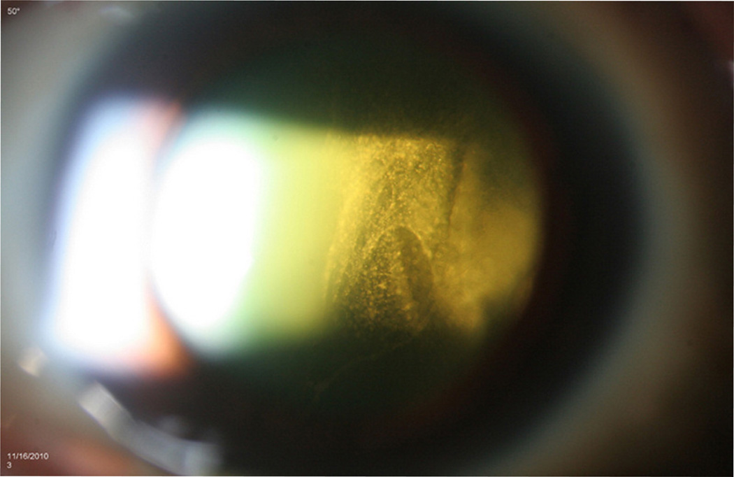
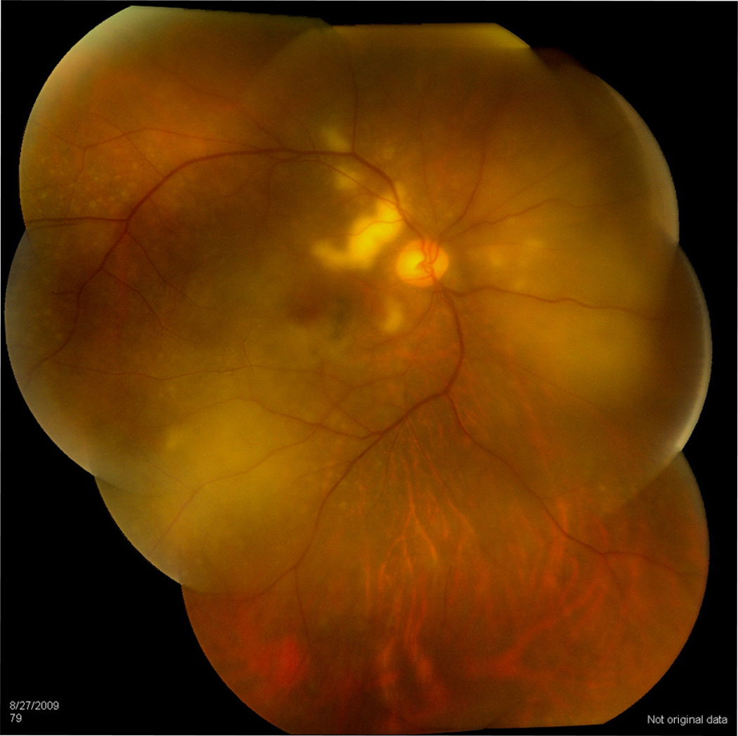
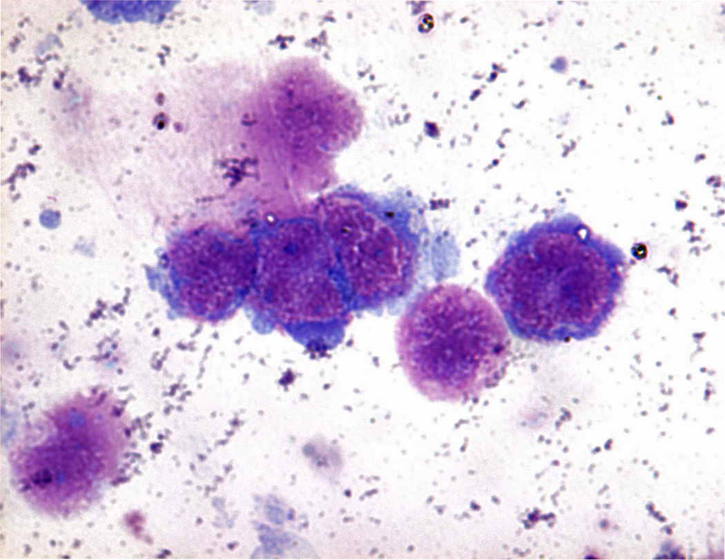
Primary vitreoretinal lymphoma (PVRL), previously called primary intraocular lymphoma (PIOL), is a rare and fatal ocular malignancy. PVRL is a subset of primary central nervous system lymphoma (PCNSL), mostly a diffuse large B-cell lymphoma. The diagnosis of PVRL is often challenging as it often masquerades as chronic uveitis. PVRL requires invasive procedures for tissue diagnosis. Cytology/pathology, molecular pathology (immuno-globulin or T-cell receptor gene rearrangement), immunohistochemistry, biophysical technology (flow cytometry), and cytokine analysis (interleukine-10) are often required. The therapies that have been successful in systemic lymphomas have not been reliably effective in PVRL and PCNSL. Current management of PVRL involves aggressive chemotherapy (methotrexate and rituximab) and radiation therapy. PVRL normally responds well to initial treatment; however, relapse rate and CNS involvement are high, resulting in poor prognosis and limited survival. A professional team of medical experts in ophthalmology, oncology (particularly neuro-oncology), and pathology is essential for optimizing patient management.
Conflict of interest statement
The authors report no conflicts of interest.
Figures
References
-
- Cassoux N, Merle-Beral H, Leblond V, Bodaghi B, Milea D, Gerber S, Fardeau C, Reux I, Xuan KH, Chan CC, Lehoang P. Ocular and central nervous system lymphoma: clinical features and diagnosis. Ocul Immunol Inflamm. 2000;8(4):243–250. - PubMed
-
- Cassoux N, Merle-Beral H, Lehoang P, Herbort C, Chan CC. Interleukin-10 and intraocular-central nervous system lymphoma. Ophthalmology. 2001;108(3):426–427. - PubMed
Publication types
MeSH terms
Grants and funding
LinkOut - more resources
Full Text Sources
Medical