. 2013 Feb 28;4(2):e520.
doi: 10.1038/cddis.2013.47.

Role of protein kinase C δ in ER stress and apoptosis induced by oxidized LDL in human vascular smooth muscle cells

Affiliations

Role of protein kinase C δ in ER stress and apoptosis induced by oxidized LDL in human vascular smooth muscle cells

P Larroque-Cardoso et al. Cell Death Dis. .

Abstract

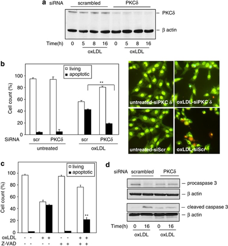
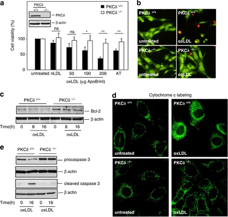
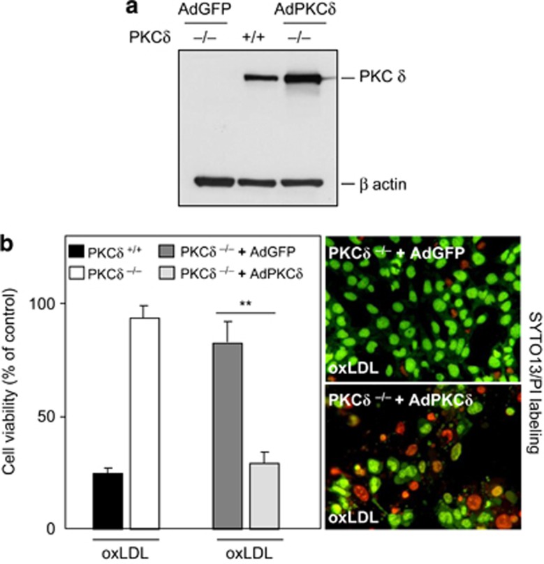
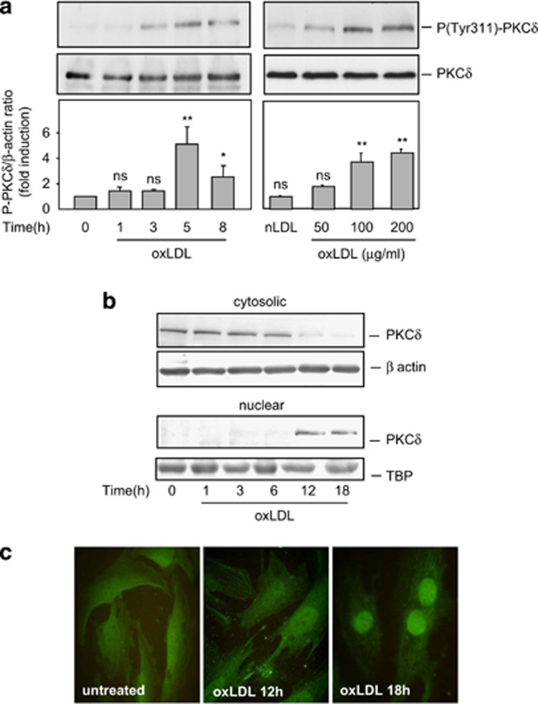
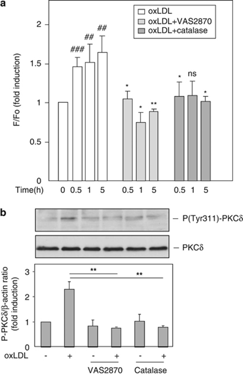
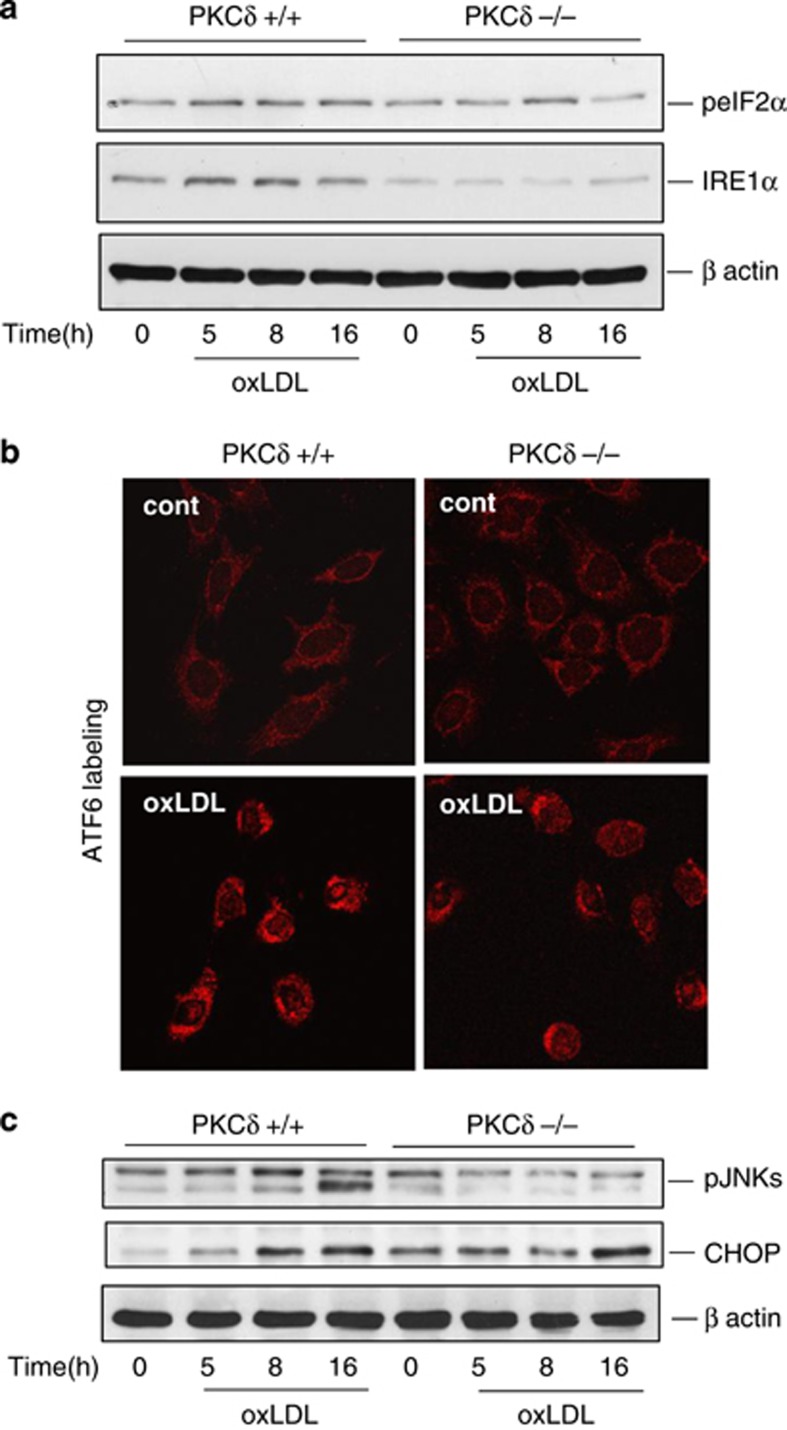
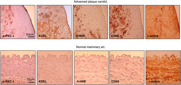
During atherogenesis, excess amounts of low-density lipoproteins (LDL) accumulate in the subendothelial space where they undergo oxidative modifications. Oxidized LDL (oxLDL) alter the fragile balance between survival and death of vascular smooth muscle cells (VSMC) thereby leading to plaque instability and finally to atherothrombotic events. As protein kinase C δ (PKCδ) is pro-apoptotic in many cell types, we investigated its potential role in the regulation of VSMC apoptosis induced by oxLDL. We found that human VSMC silenced for PKCδ exhibited a protection towards oxLDL-induced apoptosis. OxLDL triggered the activation of PKCδ as shown by its phosphorylation and nuclear translocation. PKCδ activation was dependent on the reactive oxygen species generated by oxLDL. Moreover, we demonstrated that PKCδ participates in oxLDL-induced endoplasmic reticulum (ER) stress-dependent apoptotic signaling mainly through the IRE1α/JNK pathway. Finally, the role of PKCδ in the development of atherosclerosis was supported by immunohistological analyses showing the colocalization of activated PKCδ with ER stress and lipid peroxidation markers in human atherosclerotic lesions. These findings highlight a role for PKCδ as a key regulator of oxLDL-induced ER stress-mediated apoptosis in VSMC, which may contribute to atherosclerotic plaque instability and rupture.


Figures

Figure 1
Figure 1
SiRNA silencing of PKCδ reduced oxLDL-induced apoptosis of human VSMC. (a) Representative western blot of the expression of PKCδ after siRNA silencing. Human VSMC were transfected with 100 nM scrambled siRNA or 100 nM PKCδ siRNA for 48 h as described under ‘Materials and Methods.' After siRNA transfection, hVSMC were treated with oxLDL (200 μg ApoB/ml) at the indicated times. Immunoblots were performed on cell lysates using an anti-PKCδ antibody and β-actin was used as a loading control. (b) Analysis of oxLDL-induced cell death. Human VSMC transfected with scrambled (scr) or PKCδ siRNA were incubated with oxLDL (200 μg ApoB/ml) for 24 h and apoptotic cells were counted after staining with SYTO-13/PI as described under ‘Materials and Methods.' The left graph represents the results expressed as percentage of untreated control and the mean±S.E.M. of four independent experiments (>200 cells were counted for each variable per experiment). **P<0.01 indicates significance (comparison between apoptotic cells from scr siRNA transfected cells+oxLDL and PKCδ siRNA transfected cells+oxLDL groups. The left panel illustrates the SYTO-13/IP labeling of human VSMC treated or not with oxLDL (200 μg ApoB/ml, for 24 h). (c, d) Involvement of caspase-3 in oxLDL-induced apoptosis. (c) Human VSMC pretreated with z-VAD-fmk (50 μM) were incubated with oxLDL (200 μg ApoB/ml) for 24 h and apoptotic cells were counted after staining with SYTO-13/PI as described under ‘Materials and Methods.' The graph represents the results expressed as percentage of untreated control and the mean±S.E.M. of four independent experiments (>200 cells were counted for each variable per experiment). **P<0.01 indicates significance (comparison between apoptotic cells from oxLDL-treated and z-VAD+oxLDL-treated groups). (d) Representative western blot of time-course analysis of procaspase-3 processing and cleaved caspase-3 generation in human VSMC transfected with scrambled or PKCδ siRNA and treated with oxLDL (200 μg ApoB/ml, 16 h). Immunoblots representative of three independent experiments, were performed on cell lysates using anti-procaspase-3, anti-cleaved caspase-3 antibodies and β-actin was used as a loading control
Figure 2
Figure 2
MEF PKCδ−/ are protected from oxLDL-induced apoptosis. (a) Analysis of cell toxicity in MEF PKCδ−/ and MEF wild-type (named PKCδ+/+) was evaluated by the MTT assay. MEF PKCδ and MEF PKCδ+/+ were treated with oxLDLs (50–200 μg ApoB/ml), native LDL (nLDL, 100 μg ApoB/ml) or antimycin A (10 μM) for 24 h and cell toxicity was analyzed as described. Results are expressed as percentage of untreated control and represent the mean±S.E.M. of five separate experiments. **P<0.01 and *P<0.05 indicate significance (comparison were made between PKCδ−/ and PKCδ +/+ treated with 100 and 200 oxLDL μg ApoB/ml or 10 μM antimycin A), ns indicates no significance. (b) SYTO-13/PI staining of MEF PKCδ−/ and MEF PKCδ+/+ treated or not with oxLDL (200 μg ApoB/ml, for 24 h), the images illustrate the resistance of MEF PKCδ−/ towards oxLDL-induced apoptosis. (c) Time-course analysis of Bcl-2 expression in MEF PKCδ−/ and MEF PKCδ+/+ treated with oxLDL (200 μg ApoB/ml). Immunoblots representative of three independent experiments were performed on cell lysates using anti-Bcl-2 antibody and β-actin was used as a loading control. (d) Immunocytochemistry experiments showing the release of the cytochrome C monitored by immunofluorescence in MEF PKCδ−/ and MEF PKCδ+/+ treated with oxLDL (200 μg apoB/ml) 16 h. Cells were fixed and labeled with anti-cytochrome C antibody. The results are representative of three separate experiments. (e) Representative western blot of time-course analysis of procaspase-3 processing and cleaved caspase-3 generation in MEF PKCδ−/ and MEF PKCδ+/+ treated with oxLDL (200 μg ApoB/ml, 16 h). Immunoblots representative of three independent experiments were performed on cell lysates using anti-procaspase-3, anti-cleaved caspase-3 antibodies and β-actin was used as a loading control
Figure 3
Figure 3
Re-expression of PKCδ–GFP in MEF PKCδ−/ cells reconstitutes apoptotic potential. (a) Western blot analysis of re-expression of PKCδ in MEF PKCδ−/ compared with MEF PKCδ+/+. MEF PKCδ−/ were infected with adenovirus GFP (AdGFP) or a PKCδ–GFP (AdPKCδ–GFP) fusion protein for 24 h. Immunoblots were performed on cell lysates using anti-PKCδ and β-actin was used as a loading control. (b) Analysis of cell toxicity in MEF PKCδ−/ expressing AdPKCδ–GFP or AdGFP fusion protein was evaluated by the MTT assay. MEF PKCδ−/ infected with adenovirus GFP (AdGFP) or a PKCδ–GFP (AdPKCδ–GFP) fusion protein for 24 h. Cells were treated or not with oxLDLs (200 μg ApoB/ml) for 24 h and cell toxicity (left panel) was analyzed as described. Results are expressed as percentage of untreated control and represent the mean±S.E.M. of four separate experiments. **P<0.01 indicate significance (comparison were made between PKCδ−/+AdGFP and PKCδ−/+AdPKCδ–GFP treated with oxLDL). SYTO-13/PI labeling (right panel) of PKCδ−/+AdGFP and PKCδ−/+AdPKCδ–GFP) treated with oxLDL (200 μg ApoB/ml, for 24 h), the images illustrate the restoration of the apoptotic potential of MEF PKCδ−/ expressing AdPKCδ–GFP. Cells treated with oxLDL showed chromatin condensation and appearance of apoptotic bodies, because of the brightness of SYTO-13, GFP fluorescence is not apparent
Figure 4
Figure 4
OxLDL induce PKCδ activation through tyrosine 311 phosphorylation and nuclear translocation in human VSMC. (a) Time-course analysis of PKCδ phosphorylation in human VSMC treated with oxLDL (200 μg ApoB/ml). Western blot experiments were performed on total protein extracts using anti-phosphotyrosine 311 PKCδ antibody and β-actin expression was used as loading control. The graph represents values of phosphotyrosine 311 PKCδ band intensity after normalization for total PKCδ by densitometry, *P<0.05 and **P<0.01 indicate significance (comparison between untreated cells and oxLDL 5 h and between untreated cells and oxLDL 8 h), ns indicates no significance. Blots are representative of four independent experiments. (b) Time-course analysis showing nuclear translocation of PKCδ in human VSMC treated with oxLDL (200 μg ApoB/ml). Immunoblots were performed on cell lysates and analyzed for the presence of PKCδ after nuclear and cytosolic fractionation as described under ‘Materials and Methods.' TBP (TATA-binding protein, nuclear marker) and β-actin (cytosolic marker) were also used as a loading control. Blots are representative of three independent experiments. (c) Immunocytochemistry experiments showing the nuclear translocation of PKCδ monitored by immunofluorescence in human VSMC treated with oxLDL (200 μg ApoB/ml) for 12 or 18 h. Cells were fixed and labeled with anti-PKCδ antibody. The results are representative of three separate experiments
Figure 5
Figure 5
ROS generation mediated by oxLDL is involved in the activation of PKCδ. (a) Measurement of intracellular ROS production using the free radical sensor: H2DCFDA (6-carboxy-2′,7′-dichlorodihydrofluorescein diacetate). Human VSMC were pretreated with the NADPH inhibitor (VAS-2870, 10 μM) or with PEG-catalase (50 UI/ml) for 1 h, then incubated with oxLDL (200 μg apoB/mL) for 0.5, 1 and 5 h. The variation of intracellular ROS was detected by fluorescence intensity using using the free radical sensor: 6-carboxy-2′,7′-dichlorodihydrofluorescein diacetate as described under ‘Materials and Methods.' Results were normalized on protein levels and expressed in ratio to untreated control. The data are expressed as mean±S.E.M. of five separate experiments *or #P<0.05; **or ##P<0.01 indicate significance, (# comparison between untreated cells and oxLDL-treated cells, *comparison between oxLDL-treated cells and oxLDL+VAS2870, between oxLDL-treated cells and oxLDL+catalase). ns indicates no significance. (b) Analysis of oxLDL-induced PKCδ phosphorylation in human VSMC pretreated with NADPH inhibitor (VAS-2870, 10 μM) or with PEG-catalase (50 UI/ml). Western blot experiments were performed on total protein extracts treated with oxLDL (200 μg ApoB/ml) for 8 h, using anti-phosphotyrosine 311 PKCδ antibody and total PKCδ expression was used as loading control. The graph represents values of phosphotyrosine 311 PKCδ band intensity after normalization for total PKCδ by densitometry, **P<0.01 indicates significance (comparison between oxLDL and oxLDL+VAS2870, between oxLDL and oxLDL+catalase). Blots are representative of three independent experiments
Figure 6
Figure 6
OxLDL trigger UPR and pro-apoptotic ER stress pathways in human VSMC. Time course of ER stress sensors activation in human VSMC treated with oxLDL (200 μg ApoB/ml). (a) Western blot experiments were performed on total protein extracts, cell lysates were assessed for phospho-eIF2α and IRE1α expression. β-Actin was used as protein loading control. Blots are representative of three independent experiments. (b) Immunocytochemistry experiments show the cytoplasmic and nuclear translocation of ATF6 in human VSMC treated with oxLDL (200 μg ApoB/ml) for 16 h. These data are representative of three separate experiments. (c, d) Time course of the ER stress pro-apoptotic mediators activation in human VSMC treated with oxLDL (200 μg ApoB/ml). Western blot experiments were performed on total protein extracts, cell lysates were assessed for phospho-JNK, CHOP, BIM and PUMA expression. β-Actin was used as protein loading control. Blots are representative of three independent experiments
Figure 7
Figure 7
PKCδ is involved in the activation of the pro-apoptotic ER stress IRE1/JNK pathway but not in CHOP activation. Time course of ER stress sensors activation in MEF PKCδ −/ and MEF PKCδ+/+ treated with oxLDL (200 μg ApoB/ml). (a) Western blot experiments were performed on total protein extracts, cell lysates were assessed for phospho-eIF2α and IRE1α expression. β-Actin was used as protein loading control. Blots are representative of three independent experiments. (b) Immunocytochemistry experiments show the cytoplasmic and nuclear translocation of ATF6 in MEF PKCδ−/ and MEF PKCδ+/+ treated with oxLDL (200 μg ApoB/ml) for 16 h. These data are representative of three separate experiments. (c) Time course of the ER stress proapoptotic mediators activation in MEF PKCδ−/ and MEF PKCδ+/+ treated with oxLDL (200 μg ApoB/ml). Western blot experiments were performed on total protein extracts, cell lysates were assessed for phospho-JNK and CHOP expression. β-Actin was used as protein loading control. Blots are representative of three independent experiments
Figure 8
Figure 8
PKCδ colocalized with ER stress and lipid peroxidation markers in advanced human carotid plaques. Immunostaining of human carotid plaque specimens (upper panel) and normal mammary arteries (lower panel) with anti-phosphotyrosine 311 PKCδ, anti-KDEL, anti-4-HNE-adduct, anti-CD68 and anti-α-actin antibodies. The scale bars represent 50 μm (advanced carotid plaque) and 25 μm (normal mammary artery)

References

    1. Steinberg D, Parthasarathy S, Carew TE, Khoo JC, Witztum JL. Beyond cholesterol. Modifications of low-density lipoprotein that increase its atherogenicity. N Engl J Med. 1989;320:915–924. - PubMed
    1. Napoli C. Oxidation of LDL, atherogenesis, and apoptosis. Ann NY Acad Sci. 2003;1010:698–709. - PubMed
    1. Salvayre R, Auge N, Benoist H, Negre-Salvayre A. Oxidized low-density lipoprotein-induced apoptosis. Biochim Biophys Acta. 2002;1585:213–221. - PubMed
    1. Clarke MC, Figg N, Maguire JJ, Davenport AP, Goddard M, Littlewood TD, et al. Apoptosis of vascular smooth muscle cells induces features of plaque vulnerability in atherosclerosis. Nat Med. 2006;12:1075–1080. - PubMed
    1. Ingueneau C, Huynh-Do U, Thiers JC, Negre-Salvayre A, Salvayre R, Vindis C. Caveolin-1 sensitizes vascular smooth muscle cells to mildly oxidized LDL-induced apoptosis. Biochem Biophys Res Commun. 2008;369:889–893. - PubMed

Publication types

MeSH terms