. 2013 Jan;5(1):37-50.
doi: 10.18632/aging.100527.

Specific lipofuscin staining as a novel biomarker to detect replicative and stress-induced senescence. A method applicable in cryo-preserved and archival tissues

Affiliations

Specific lipofuscin staining as a novel biomarker to detect replicative and stress-induced senescence. A method applicable in cryo-preserved and archival tissues

E A Georgakopoulou et al. Aging (Albany NY). 2013 Jan.

Abstract

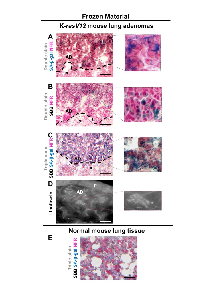
There is shortage of extensive clinicopathologic studies of cellular senescence because the most reliable senescence biomarker, the detection of Senescence-Associated-beta-galactosidase activity (SA-β-gal), is inapplicable in archival material and requires snap-frozen tissues. We validated the histochemical Sudan-Black-B (SBB) specific stain of lipofuscin, an aggregate of oxidized proteins, lipids and metals, known to accumulate in aged tissues, as an additional reliable approach to detect senescent cells independently of sample preparation. We analyzed cellular systems in which senescence was triggered by replicative exhaustion or stressful stimuli, conditional knock-in mice producing precancerous lesions exhibiting senescence, and human preneoplastic lesions known to contain senescent cells. In the above settings we demonstrated co-localization of lipofuscin and SA-β-gal in senescent cells in vitro and in vivo (cryo-preserved tissue), strongly supporting the candidacy of lipofuscin for a biomarker of cellular senescence. Furthermore, cryo-preserved tissues positive for SA-β-gal were formalin-fixed, paraffin-embedded, and stained with SBB. The corresponding SA-β-gal positive tissue areas stained specifically for lipofuscin by SBB, whereas tissues negative for SA-β-gal were lipofuscin negative, validating the sensitivity and specificity of the SBB staining to visualize senescent cells in archival material. The latter unique property of SBB could be exploited in research on widely available retrospective tissue material.


Conflict of interest statement

The authors of this manuscript have no conflict of interests to declare.

Figures

Figure 1
Figure 1. Lipofuscin accumulates and co-localizes with Senescence-Associated beta-galactosidase (SA-β-gal) in sub-confluent senescent primary human diploid lung fibroblasts (DLF)
Y (Young): Early- passage cells, RS: Replicative-senescent cells and IS (irradiated): Early passage cells that became prematurely senescent after irradiation (12×4Gy). Collected cells were fixed on slides with 4% parafolmadehyde (A) All three cultures were stained with SA-β-gal and nuclear fast red as counterstain (NFR). Cells from RS and IS cultures acquired the characteristic senescent morphological phenotype (enlarged and flattened) and were positive for SA-β-gal staining (turquoise color). (B) All cultures were stained with Sudan Black B (SBB) and NFR. Cells from RS and IS cultures, which had the morphological phenotype of senescence, were also positive for SBB (dark blue-black granules). (C) Top panels: green pseudocolor represents visualization of lipofuscin's autofluorescence at 450-490 nm. Bottom panels: RS and IS cells that stained with SBB (BF, bright field microscopy) show no auto-fluorescence of lipofuscin (FM, fluorescence microscopy without pseudocolor), indicating that SBB stains lipofuscin. Cells with the morphological phenotype of senescence were positive for both SA-β-gal and SBB (D), while cells that were positive for Ki67 were negative for both SA-β-gal and SBB (E). Insets: Cells at higher magnification, pink dashed lines: indicate NFR-stained nuclei, brown dashed lines: indicate Ki67- negative nuclei, black arrows: show SBB granules.
Figure 2
Figure 2. Lipofuscin accumulates and co-localizes with Senescence-Associated beta-galactosidase (SA-β-gal) in senescent Saos-2 cells triggered by p21WAF-1
(A) SA-β-gal staining (turquoise color) in the Saos-2 p21WAF-1 Tet-On cell system on the 8th day of doxycycline (5 μg/ml) addition. Inset: Senescent cells acquired the characteristic senescent morphological phenotype (enlarged and flattened). (B) Sudan Black B (SBB) positivity (dark blue-black granules) in cells with senescent morphological phenotype (inset). (C) Top panels: Lipofuscin's auto-fluorescence in induced Saos-2 p21WAF-1 Tet-On cells, by fluorescence microscopy at 450-490 nm (green pseudocolor). Bottom panels: Cytochemical SBB staining (BF, bright field microscopy) quenches the auto-fluorescence of lipofuscin (FM, fluorescence microscopy), indicating that SBB stains lipofuscin. SA-β-gal and SBB staining coincided in cells that had the morphological phenotype of senescence (D) and were absent in cells that were positive for the proliferative marker Ki67 (E). (F) Addition of doxycyclin (Dox) triggers p21WAF-1 expression. Brown dashed lines: Ki67- negative nuclei. Black arrows: SBB granules. NFR: nuclear fast red counterstain.
Figure 3
Figure 3. Lipofuscin accumulates and co-localizes with Senescence-Associated beta-galactosidase (SA-β-gal) in p53-mediated Saos-2 senescent cells
(A) Senescent cells with the characteristic morphology (enlarged and flattened) and positivity for SA-β-gal staining (turquoise color), on the 8th day of induced with doxycycline of the Saos-2 p53 Tet-On system. (B) Sudan Black B (SBB) perinuclear accumulation as dark blue-black granules, in cells with senescent morphology. (C) Top panels: Perinuclear appearance of lipofuscin in apparently senescent cells: pseudocolor visualization of lipofuscin's auto-fluorescence (450-490 nm) is represented in green. Bottom panels: Lipofuscin's auto-fluorescence (FM, fluorescence microscopy) is masked by SBB staining (BF, bright field microscopy). (D) Co-localization of SA-β-gal and SBB staining in senescent cells and, (E) Ki67 positive cells are negative for SBB and SA-β-gal. (F) Addition of 5μg/ml doxycyclin (Dox) leads to p53 expression. Brown dashed lines: Ki67- negative nuclei, black arrows: SBB granules, NFR: nuclear fast red counterstain.
Figure 4
Figure 4. Lipofuscin accumulates and co-localizes with Senescence-Associated beta-galactosidase (SA-β-gal) in E2F-1 induced U2OS senescent cells
(A) On the 10th day of induction with 4-OH-Tamoxifen, cells were positive for SA-β-gal activity (turquoise color); cells also demonstrated the morphological phenotype of senescence (enlarged and flattened) (B) Cells demonstrating the characteristic senescent phenotype show Sudan Black B (SBB) dark blue-black granules (C) Top panels: Lipofuscin's auto-fluorescence at 450-490 nm is represented in green pseudocolor. Bottom panels: blocking of lipofuscin auto-fluorescence (FM, fluorescence microscopy) with SBB staining (BF, bright field microscopy) indicates that SBB stains lipofuscin (D) Concurrent positivity for SA-β-gal activity and SBB staining in the same cell, which is also negative for the proliferative marker Ki67 (E). (F) Addition of 300 nmol/L 4-OH-Tamoxifen (4-OH-Tam) leads to nuclear translocation of E2F1 (indirect immunofluorescence). E2F1-negative nuclei are indicated with white dashed lines. Brown dashed lines: Ki67- negative nuclei, black arrows: SBB granules, NFR: nuclear fast red counterstain.
Figure 5
Figure 5. Lipofuscin and Senescence-Associated beta-galactosidase (SA-β-gal) activity co-localize in lung adenomas demonstrating senescence in a mouse model conditionally expressing K-rasV12 in the lung
Frozen sections derived from mouse lung K-rasV12 adenomas. (A) Cells from the adenomas show SA-β-gal activity. (B) Characteristic perinuclear deposition of blue black granules in cells stained with Sudan Black B (SBB), representing positivity for lipofuscin. (C) Cells from lung adenomas positive for both, SA-β-gal activity and lipofuscin. (D) Fluorescence microscopy (at 450-490 nm) verifying lipofuscin presence. (E) Normal mouse lung tissue negative for SA-β-gal activity and lipofuscin. P: Parenchyma, AD: Adenoma. Scale bars: A-C, 200 μm; D, 25 μm; E, 50 μm. Insets: Cells at higher magnification.
Figure 6
Figure 6. Lipofuscin accumulates and co-localizes with Senescence-Associated beta-galactosidase (SA-β-gal) in senescent cells detected in cryo-preserved material from benign prostatic hyperplasia (BPH)
Frozen material from patients with BPH in enlarged prostates (prostate weight greater than 55gr) was thin-sectioned (5 μm). The sections were immediately double stained for SA-β-gal activity (turquoise color) and Nuclear Fast Red (NFR) as counterstain (A) and double stained with SBB and NFR (B). Areas with characteristic BPH pathology showed SA-β-gal activity and lipofuscin positivity (C). Normal prostate regions adjacent to BPH, were found negative for SA-β-gal activity and lipofuscin (D). Scale bars: A-C, 100 μm; D, 50 μm. Insets: Cells at higher magnification.
Figure 7
Figure 7. Sudan Black B (SBB) staining demonstrates lipofuscin accumulation in lung adenomas (AD) and absence in adenocarcinomas (AdCa), in formalin-fixed paraffin-embedded (FFPE) lung sections from mice conditionally expressing K-rasV12
(A) Haematoxylin and Eosin staining demonstrates a lung adeoma (AD) and an adenocarcinoma (AdCa) on the same FFPE section (B) Histological features of the adenoma and the adenocarcinoma shown in A section. (C) Characteristic perinuclear deposition of blue black granules in adenoma cells stained with Sudan Black B (SBB), representing positivity for lipofuscin, while adenocarcinoma cells are negative for SBB. (D) Fluorescence microscopy (at 450-490 nm) verifying lipofuscin presence in the adenoma, and absence in the adenocarcinoma. Scale bars: A, 600 μm; B-D, 100 μm. Inset: Cells at higher magnification.
Figure 8
Figure 8. Accumulation of lipofuscin in formalin-fixed paraffin-embedded (FFPE) tissues from benign prostatic hyperplasia (BPH) that corresponds to senescent areas as depicted by Senescence-Associated beta-galactosidase (SA-β-gal) in cryo-preserved material
FFPE sections from patients with BPH in enlarged prostates (prostate weight greater than 55gr) demonstrate accumulation of lipofuscin. Sections were deparaffinized and double stained with SBB (dark blue-black granules) and NFR as counterstain (A). Lipofuscin's presence was verified with fluorescence microscopy (B). Immunostaining for Ki-67 shows no matching with lipofuscin accumulation (C). Normal prostate regions adjacent to BPH, were negative for lipofuscin (D). Scale bars: BPH, 100 μm; Normal Prostate, 50 μm. Insets: Cells at higher magnification.
Figure 9
Figure 9. Co-localization of Senescence-Associated beta-galactosidase (SA-β-gal) activity and lipofuscin depiction in fresh-frozen tissue sample of benign prostatic hyperplasia (BPH) pretreated with SA-β-gal and subsequently embedded in paraffin
Fresh samples with BPH were snap frozen, fixed in 4% formaldehyde, washed with buffer, incubated in SA-β-gal solution (turquoise color), subsequently fixed with formal-dehyde, and then embedded in paraffin, as previously shown (Michaloglou et al 2005). Sections where then double stained with Sudan Black B (SBB) (dark blue-black granules) and Nuclear Fast Red (NFR) as counterstain. Areas with the characteristic pathology of BPH showed SA-β-gal activity and lipofuscin positivity. Note the weak intensity of the Sa-β-gal staining.
Figure 10
Figure 10. Lipofuscin staining and Senescence-Associated beta-galactosidase (SA-β-gal) activity in primary human diploid lung fibroblasts (DLF)
Triple staining of early passage (6th) DLF cells with Sudan Black B (SBB) (dark blue-black granules), SA-β-gal (turquoise color) and Nuclear Fast Red (NFR), as counterstain. (A) Cells that had just reached 100% confluence showed SA-β-gal activity with negligible lipofuscin (arrows). (B) In the same assay 72 hours later, some cells demonstrated SA-β-gal staining (arrowheads) without lipofuscin, and there were also cells with concurrent SA-β-gal and SBB staining (arrows). (C) In sub-confluent DLF cells, prolonged SA-β-gal incubation (72 hours) followed by immediate SBB staining, demonstrated SA-β-gal activity (inset) without lipofuscin appearance.

References

    1. Gorgoulis VG, Halazonetis T. Oncogene-induced senescence: the bright and dark side of the response. Curr Opin Cell Biol. 2010;22:816–827. - PubMed
    1. Dimri GP, Lee X, Basile G, Acosta M, Scott G, Roskelley C, Medrano EE, Linskens M, Rubelj I, Pereira-Smith O, Peacocke M, Campisis J. A biomarker that identifies senescent human cells in culture and in aging skin in vivo. Proc Natl Acad Sci U S A. 1995;92:9363–9367. - PMC - PubMed
    1. Sikora E, Arendt T, Bennett M, Narita M. Impact of cellular senescence signature on ageing research. Ageing Res Rev. 2011;10:146–152. - PubMed
    1. Hayflick L. The limited in vitro lifespan of human diploid cell strains. Exp Cell Res. 1965:614–636. - PubMed
    1. Collado M, Gil J, Efeyan A, Guerra C, Schuhmacher AJ, Barradas M, Benguría A, Zaballos A, Flores JM, Barbacid M, Beach D, Serrano M. Tumour biology: senescence in premalignant tumours. Nature. 2005;436:642. - PubMed

Publication types