

Prion infectivity in the spleen of a PRNP heterozygous individual with subclinical variant Creutzfeldt-Jakob disease
- PMID: 23449776
- PMCID: PMC3613713
- DOI: 10.1093/brain/awt032
Prion infectivity in the spleen of a PRNP heterozygous individual with subclinical variant Creutzfeldt-Jakob disease
Abstract
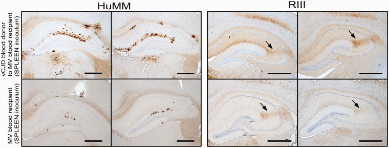
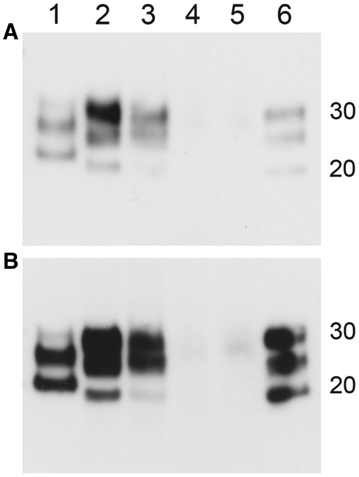
Blood transfusion has been identified as a source of human-to-human transmission of variant Creutzfeldt-Jakob disease. Three cases of variant Creutzfeldt-Jakob disease have been identified following red cell transfusions from donors who subsequently developed variant Creutzfeldt-Jakob disease and an asymptomatic red cell transfusion recipient, who did not die of variant Creutzfeldt-Jakob disease, has been identified with prion protein deposition in the spleen and a lymph node, but not the brain. This individual was heterozygous (MV) at codon 129 of the prion protein gene (PRNP), whereas all previous definite and probable cases of variant Creutzfeldt-Jakob disease have been methionine homozygotes (MM). A critical question for public health is whether the prion protein deposition reported in peripheral tissues from this MV individual correlates with infectivity. Additionally it is important to establish whether the PRNP codon 129 genotype has influenced the transmission characteristics of the infectious agent. Brain and spleen from the MV blood recipient were inoculated into murine strains that have consistently demonstrated transmission of the variant Creutzfeldt-Jakob disease agent. Mice were assessed for clinical and pathological signs of disease and transmission data were compared with other transmission studies in variant Creutzfeldt-Jakob disease, including those on the spleen and brain of the donor to the index case. Transmission of variant Creutzfeldt-Jakob disease was observed from the MV blood recipient spleen, but not from the brain, whereas there was transmission from both spleen and brain tissues from the red blood cell donor. Longer incubation times were observed for the blood donor spleen inoculum compared with the blood donor brain inoculum, suggesting lower titres of infectivity in the spleen. The distribution of vacuolar pathology and abnormal prion protein in infected mice were similar following inoculation with both donor and recipient spleen homogenates, providing initial evidence of similar transmission properties after propagation in PRNP codon 129 MV and MM individuals. These studies demonstrate that spleen tissue from a PRNP MV genotype individual can propagate the variant Creutzfeldt-Jakob disease agent and that the infectious agent can be present in the spleen without CNS involvement.
Figures


References
-
- Bishop MT, Hart P, Aitchison L, Baybutt HN, Plinston C, Thomson V, et al. Predicting susceptibility and incubation time of human-to-human transmission of vCJD. Lancet Neurol. 2006;5:393–8. - PubMed
-
- Brown DA, Bruce ME, Fraser JR. Comparison of the neuropathological characteristics of bovine spongiform encephalopathy (BSE) and variant Creutzfeldt-Jakob disease (vCJD) in mice. Neuropathol Appl Neurobiol. 2003;29:262–72. - PubMed
-
- Bruce ME, Boyle A, McConnel I. TSE strain typing in mice. In: Lehmann S, Grassi J, editors. Techniques in prion research. Birkhauser Verlag. 2004:132–46.
-
- Bruce ME, McConnell I, Will RG, Ironside JW. Detection of variant Creutzfeldt-Jakob disease infectivity in extraneural tissues. Lancet. 2001;358:208–9. - PubMed
-
- Bruce ME, Will RG, Ironside JW, McConnell I, Drummond D, Suttie A, et al. Transmissions to mice indicate that ‘new variant' CJD is caused by the BSE agent. Nature. 1997;389:498–501. - PubMed
Publication types
MeSH terms
Substances
Grants and funding
- BBS/E/D/20251967/BB_/Biotechnology and Biological Sciences Research Council/United Kingdom
- BBS/E/D/20251968/BB_/Biotechnology and Biological Sciences Research Council/United Kingdom
- G0700640/MRC_/Medical Research Council/United Kingdom
- G0900580/MRC_/Medical Research Council/United Kingdom
- G1100616/MRC_/Medical Research Council/United Kingdom
LinkOut - more resources
Full Text Sources
Other Literature Sources
Medical
