. 2013 May;87(9):5141-50.
doi: 10.1128/JVI.03347-12. Epub 2013 Feb 28.

A systems biology examination of the human female genital tract shows compartmentalization of immune factor expression

Affiliations

A systems biology examination of the human female genital tract shows compartmentalization of immune factor expression

Adam Burgener et al. J Virol. 2013 May.

Abstract

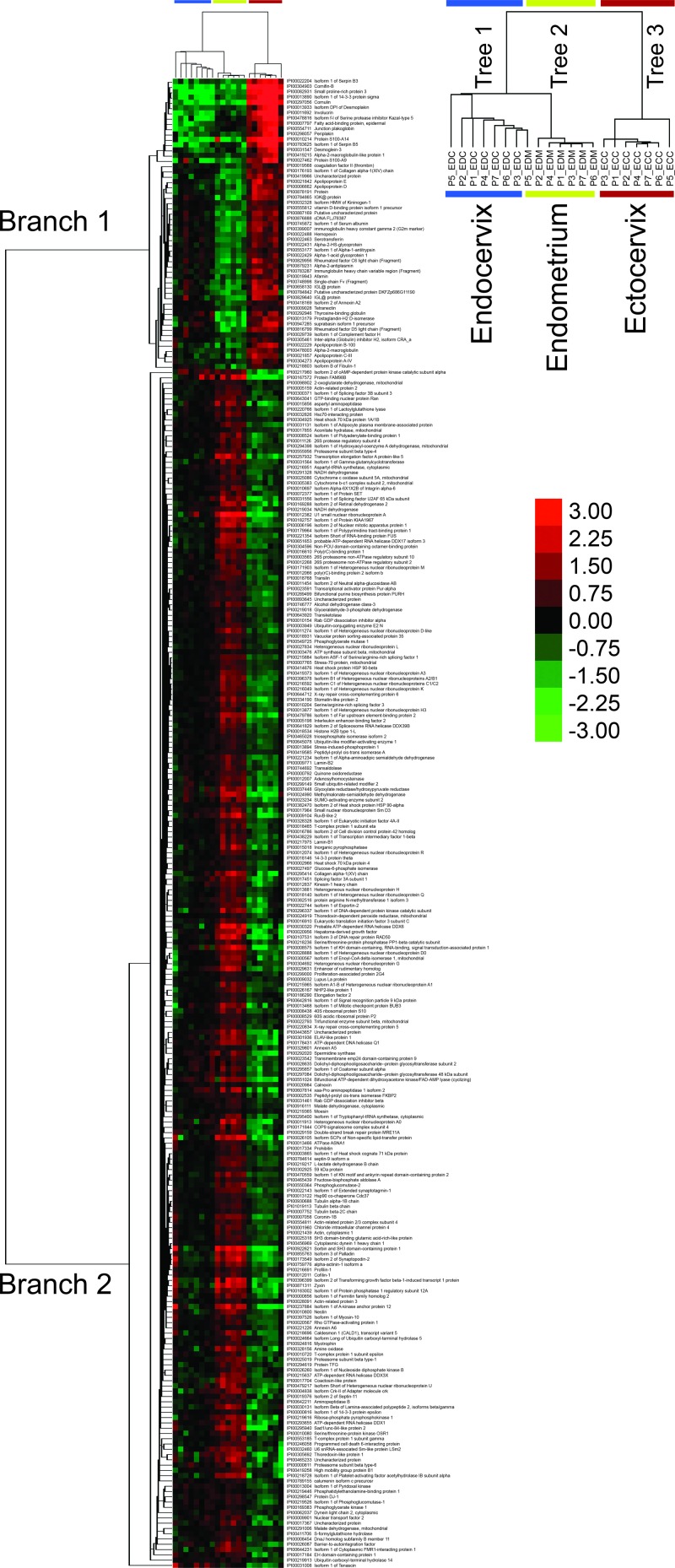
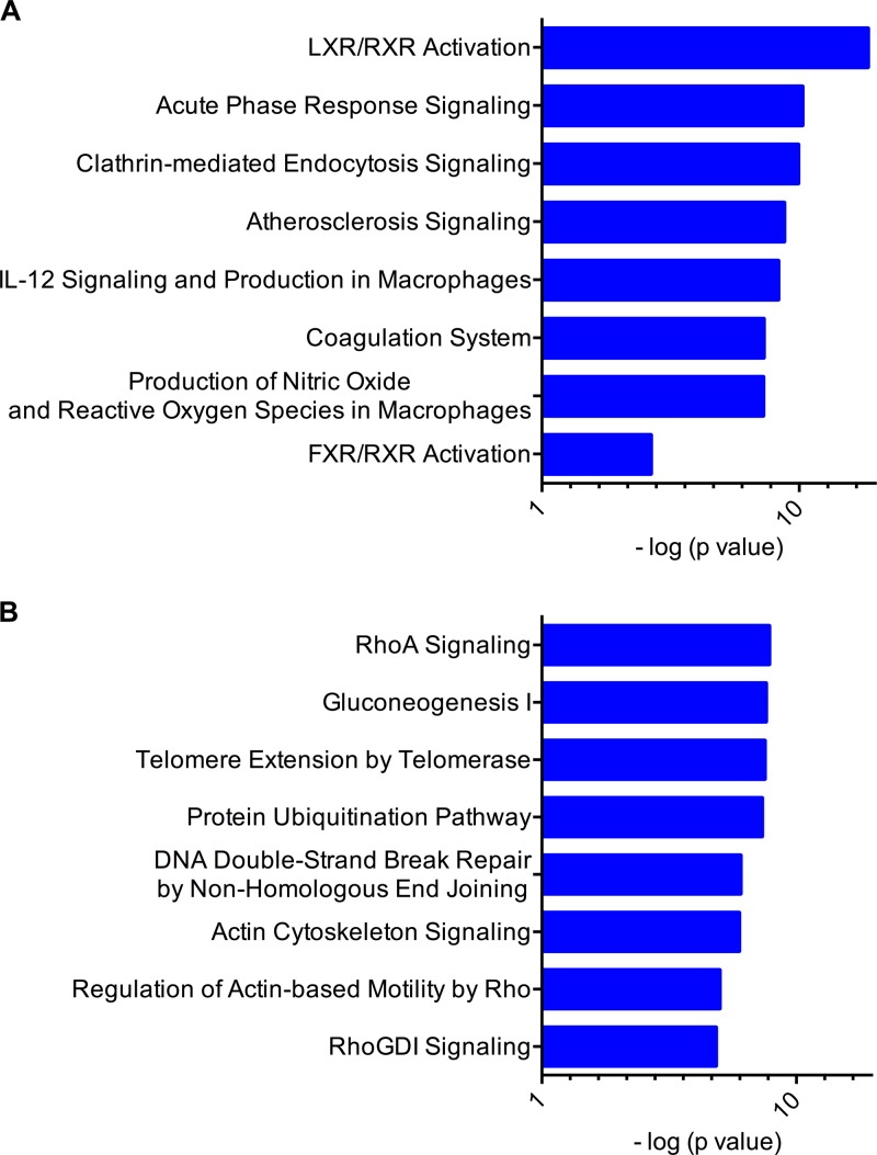
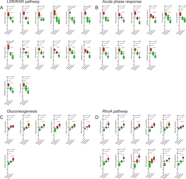
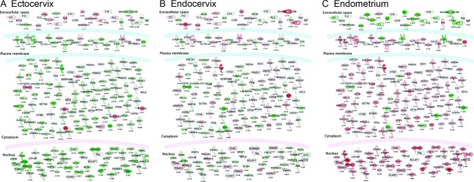
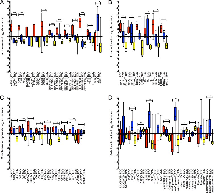
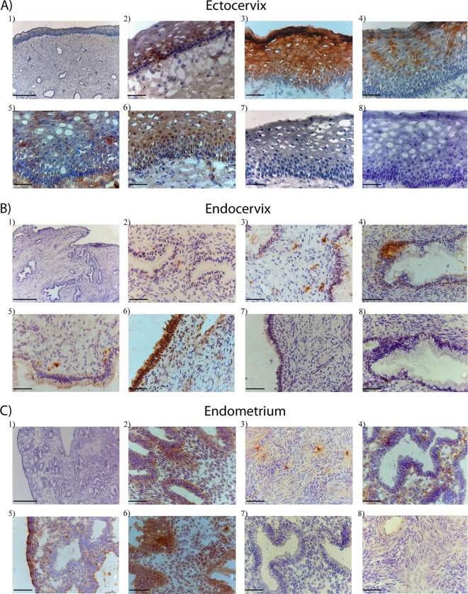
Many mucosal factors in the female genital tract (FGT) have been associated with HIV susceptibility, but little is known about their anatomical distribution in the FGT compartments. This study comprehensively characterized global immune factor expression in different tissue sites of the lower and upper FGT by using a systems biology approach. Tissue sections from the ectocervix, endocervix, and endometrium from seven women who underwent hysterectomy were analyzed by a combination of quantitative mass spectrometry and immunohistochemical staining. Of the >1,000 proteins identified, 281 were found to be differentially abundant in different tissue sites. Hierarchical clustering identified four major functional pathways distinguishing compartments, including innate immune pathways (acute-phase response, LXR/RXR) and development (RhoA signaling, gluconeogenesis), which were enriched in the ectocervix/endocervix and endometrium, respectively. Immune factors important for HIV susceptibility, including antiproteases, immunoglobulins, complement components, and antimicrobial factors, were most abundant in the ectocervix/endocervix, while the endometrium had a greater abundance of certain factors that promote HIV replication. Immune factor abundance is heterogeneous throughout the FGT and shows unique immune microenvironments for HIV based on the exposure site. This may have important implications for early events in HIV transmission and site-specific susceptibility to HIV in the FGT.


Figures

Fig 1
Fig 1
Protein abundance patterns differentiate anatomical sites of the lower and upper FGT. The heat map shown illustrates 281 proteins that were differentially abundant across tissue subsets according to one-way ANOVA (P < 1 × 10⁻⁴). Clustering of proteins and tissues was generated by unsupervised centroid linkage hierarchical clustering using the Pearson correlation coefficient as the distance metric. Protein abundance levels are shown in color, with red indicating overabundant proteins and green indicating underabundant proteins compared to the mean of all tissue samples. FGT compartment tissues (columns) are grouped together and distinguished by two distinct branches (left side) of protein abundance patterns (ECC, ectocervix; EDC, endocervix; EDM, endometrium). The heat map shows almost perfect clustering of tissue compartments based on protein abundance patterns (with the exception of EDM5, which clustered with the endocervical group).
Fig 2
Fig 2
The ectocervix and endocervix are distinguished by increased activation of immune response pathways. The top two branches identified by hierarchical clustering were analyzed by comparative IPA software. The major canonical pathways represented in branches are shown (A, branch 1; B, branch 2), with the most statistically significantly associated pathways shown in decreasing order (top to bottom). Branch 1 identifies the LXR/RXR and acute-phase response pathways as the top pathways associated with proteins found to be overabundant in ectocervical tissue. The RhoA and gluconeogenesis pathways are identified as the top pathways associated with proteins found to be overabundant in endometrial tissue. Associations calculated by a right-tailed Fisher exact test (with Benjamini-Hochberg correction) (log P values are on the horizontal axis) were used to calculate the P value of the probability that the association between each protein appearing in the data set and a canonical pathway is random. Only the top eight associated pathways are shown for vertical sizing.
Fig 3
Fig 3
Expression profiles of proteins involved in innate immunity and tissue development pathways that distinguish FGT compartments. Shown are expression profiles of proteins identified in canonical pathways most significantly associated with each branch of the hierarchical clustering analysis that distinguished FGT compartments. These include the LXR/RXR (A), acute-phase response (B), gluconeogenesis (C), and RhoA (D) pathways. The y axis represents their log2 abundance profiles (mean ± standard error of the mean) based on the FGT compartment. Colors indicate whether the average abundance in each compartment (ectocervix, endocervix, endometrium) was above (red) or below (green) the mean. Significance values across all groups were calculated by one-way ANOVA, and intergroup variations were calculated by Tukey's multiple-comparison t test (P < 0.01, *; P < 0.001, **; P < 0.0001, ***).
Fig 4
Fig 4
Cellular pathways involved in viral infectivity are differentially enriched in FGT tissue compartments. The highest-scoring immune pathways associated with differential protein abundance patterns in FGT compartments were those involved with viral infectivity according to the IPA biological function database. Proteins involved in the viral infectivity pathway are represented for each compartment (A, ectocervix; B, endocervix; C, endometrium), with the abundance log2 ratios indicated by intensity of color (green, underexpressed; red, overexpressed [compared to the average]), and their subcellular locations are shown. The endometrium shows a clear overabundance of intracellular proteins that are involved in this pathway, and the ectocervix/endocervix show a greater abundance of extracellular immune factors, such as immunoglobulins, complement components, and serpins. All of the proteins shown in color have a calculated P value of <0.05 between compartments by one-way ANOVA, and the effect of multiple comparisons was corrected for by Benjamini-Hochberg correction. Proteins in white were present in the data set but did not reach the statistical significance threshold.
Fig 5
Fig 5
Soluble immune factors are predominant in the ectocervix/endocervix and lowest in the endometrium. Shown are log2 abundance values of antiproteases (A), immunoglobulins (B), complement components (C), and antimicrobial factors (D) in FGT compartments. Compartment-specific expression is shown by color as follows: red, ectocervix (ECC); blue, endocervix (EDC); yellow, endometrium (EDM). Significance values across all groups were calculated by one-way ANOVA (P < 0.01, *; P < 0.001, **; P < 0.0001, ***).
Fig 6
Fig 6
Bright-field images of in situ staining of selected antiproteases and immunoglobulins in tissue sections from the ectocervix (A), endocervix (B), and endometrium (C). The tissue sections shown were stained with hematoxylin (blue) for visualization of cell nuclei. Images A1, B1, and C1 are ×5 overview pictures of representative ectocervix, endocervix, and endometrium tissue samples showing the epithelium and the submucosal layer. For visualization of immune proteins, tissue sections were stained (brown) for A2ML1 (A2, B2, and C2), cystatin B (A3, B3, and C3), elafin (A4, B4, and C4), IgA (A5, B5, and C5), serpin A1 (A6, B6, and C6), rabbit anti-mouse IgG (A7, B7, and C7), and rabbit anti-goat IgG (A8, B8, and C8) antibodies. The A1, B1, and C1 images were collected with a 5×/0.12 objective (scale bars, 500 μm), and all of the other images were collected with a 40×/0.65 objective (scale bars, 50 μm).

Similar articles

Cited by

References

    1. UNAIDS 2011. Global report: UNAIDS report on the global AIDS epidemic, 2010. Joint United Nations Programme on HIV/AIDS, New York, NY: http://www.unaids.org/documents/20101123_globalreport_em.pdf
    1. Valore EV, Park CH, Igreti SL, Ganz T. 2002. Antimicrobial components of vaginal fluid. Am. J. Obstet. Gynecol. 187:561–568 - PubMed
    1. Wira CR, Patel MV, Ghosh M, Mukura L, Fahey JV. 2011. Innate immunity in the human female reproductive tract: endocrine regulation of endogenous antimicrobial protection against HIV and other sexually transmitted infections. Am. J. Reprod. Immunol. 65:196–211 - PMC - PubMed
    1. Ghosh M, Fahey JV, Shen Z, Lahey T, Cu-Uvin S, Wu Z, Mayer K, Wright PF, Kappes JC, Ochsenbauer C, Wira CR. 2010. Anti-HIV activity in cervical-vaginal secretions from HIV-positive and -negative women correlate with innate antimicrobial levels and IgG antibodies. PLoS One 5:e11366 doi:10.1371/journal.pone.0011366 - DOI - PMC - PubMed
    1. Hur EM, Patel SN, Shimizu S, Rao DS, Gnanapragasam PN, An DS, Yang L, Baltimore D. 2012. Inhibitory effect of HIV-specific neutralizing IgA on mucosal transmission of HIV in humanized mice. Blood 120:4571–4582 - PMC - PubMed

Publication types

LinkOut - more resources