. 2013 May;87(9):5182-92.
doi: 10.1128/JVI.00100-13. Epub 2013 Feb 28.

Functional analysis of the murine coronavirus genomic RNA packaging signal

Affiliations

Functional analysis of the murine coronavirus genomic RNA packaging signal

Lili Kuo et al. J Virol. 2013 May.

Abstract

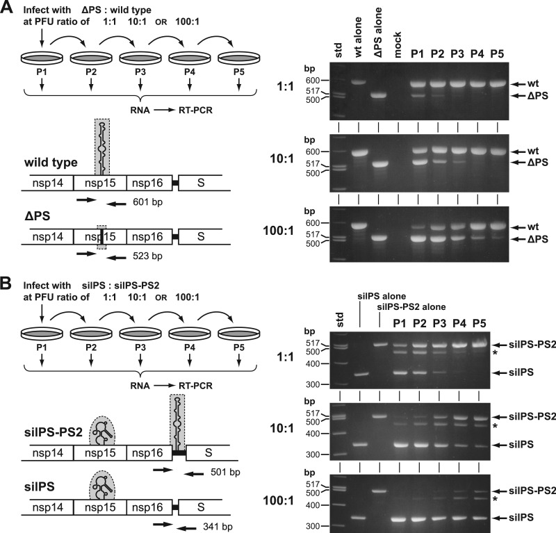
Coronaviruses selectively package genomic RNA into assembled virions, despite the great molar excess of subgenomic RNA species that is present in infected cells. The genomic packaging signal (PS) for the coronavirus mouse hepatitis virus (MHV) was originally identified as an element that conferred packaging capability to defective interfering RNAs. The MHV PS is an RNA structure that maps to the region of the replicase gene encoding the nonstructural protein 15 subunit of the viral replicase-transcriptase complex. To begin to understand the role and mechanism of action of the MHV PS in its native genomic locus, we constructed viral mutants in which this cis-acting element was altered, deleted, or transposed. Our results demonstrated that the PS is pivotal in the selection of viral genomic RNA for incorporation into virions. Mutants in which PS RNA secondary structure was disrupted or entirely ablated packaged large quantities of subgenomic RNAs, in addition to genomic RNA. Moreover, the PS retained its function when displaced to an ectopic site in the genome. Surprisingly, the PS was not essential for MHV viability, nor did its elimination have a severe effect on viral growth. However, the PS was found to provide a distinct selective advantage to MHV. Viruses containing the PS readily outcompeted their otherwise isogenic counterparts lacking the PS.


Figures

Fig 1
Fig 1
MHV RNA species. The 31.3-kb MHV genome (gRNA) is shown with an expanded segment detailing the 3′ terminus of the replicase gene (rep 1a and 1b), including the region encoding nsp14 to nsp16. The position of the packaging signal (PS) within the coding region for nsp15 is indicated. Downstream of the replicase gene are the genes for structural proteins (spike [S], envelope [E], membrane [M], and nucleocapsid [N]) and for accessory proteins (2a, hemagglutinin-esterase [HE], 4, 5a, and internal [I]). Beneath the gRNA is the 3′-nested set of transcribed subgenomic RNA (sgRNA) species that is a defining characteristic of coronaviruses and other members of the order Nidovirales.
Fig 2
Fig 2
Donor RNA transcription vector for construction of MHV PS mutants by targeted RNA recombination (22, 23). All mutants in the current work originated with the parent vector pPM9, which was derived from the previously described pMH54 (23) by the addition of genomic cDNA upstream of the S gene. The locus containing the PS is indicated by a bar above nsp15. Shown are coding-silent unique XbaI and BspEI sites flanking the PS and unique SalI and AscI sites in the truncated nonessential intergenic region between nsp16 and the S gene.
Fig 3
Fig 3
Construction of the silPS mutant. (A) Model for the MHV PS proposed by Chen et al. (17). The four repeating units with AA (or GA) bulges are boxed. (B) Positions and identities of 20 mutations (circled nucleotides) made to disrupt the structure of the PS without altering the encoded amino acid sequence of nsp15. The resulting lowest-free-energy structure predicted by Mfold (29) for the mutated sequence is shown on the right. (C) Schematics of the relevant genomic regions of the constructed silPS mutant and its isogenic wild-type counterpart. (D) Detail of the region downstream of the replicase gene, in which TRS2 was knocked out, all of gene 2a was deleted, and all except 99 nucleotides of the HE gene was deleted. The positions of SalI and AscI sites created in the parent transcription vector pPM9 are shown. Circles above the sequence indicate nucleotides that were mutated to knock out TRS2.
Fig 4
Fig 4
Growth characteristics of the silPS mutant. (A) Plaques of the silPS mutant at 33, 37, and 39°C compared with those of the isogenic wild-type virus. Plaque titrations were carried out on L2 cells; monolayers were stained with neutral red at 72 h postinfection and were photographed 18 h later. (B and C) Growth kinetics of the silPS mutant relative to the wild type. Confluent monolayers of 17Cl1 cells were infected at the indicated multiplicity of infection (moi). At the indicated times postinfection, aliquots of medium were removed, and infectious titers were determined by plaque assay on L2 cells.
Fig 5
Fig 5
RNA packaging phenotype of the silPS mutant. (A) Outline of the procedures for purification, normalization, and analysis of silPS and wild-type virions, as detailed in Materials and Methods. Infectious titers were determined by plaque assay on L2 cells at 37°C; chemiluminescence values are given in arbitrary volume units, as measured with a Bio-Rad ChemiDoc XRS+ instrument. (B) Western blot of normalized amounts of immunopurified wild-type and silPS virions probed with anti-N monoclonal antibody J.3.3 and anti-M monoclonal antibody J.1.3. (C) Coomassie blue-stained SDS-polyacrylamide gel of normalized amounts of immunopurified wild-type and silPS virions; samples on the right are a 5-fold dilution of those on the left. Molecular mass standards are indicated on the left of each panel (B and C). (D) Northern blot of total RNA isolated from infected 17Cl1 cells, from which wild-type or silPS virions were purified, or from mock-infected cells. (E) Northern blot of RNA isolated from normalized amounts of immunopurified wild-type and silPS virions. MHV RNA was detected with a probe specific for the 3′ end of the genome (D and E).
Fig 6
Fig 6
Construction of the ΔPS and ΔPS-PS2 mutants. (A) Ribbon diagram and molecular surface rendering of the MHV nsp15 monomer (PDB accession code 2GTH) (33) generated with PyMol (http://pymol.org). The nsp15 amino and carboxy termini are labeled N and C, respectively. The dotted line represents part of the surface loop connecting the amino- and carboxy-terminal domains of the protein. (B) Alignment of the central region of nsp15 of MHV with that of representative coronaviruses: lineage A betacoronaviruses BCoV and HCoV-HKU1, lineage B betacoronavirus SARS-CoV, alphacoronavirus TGEV, and gammacoronavirus IBV. GenBank accession numbers for the sequences shown are as follows: MHV, AY700211; BCoV, U00735; HCoV-HKU1, AY597011; SARS-CoV, AY278741; TGEV, AJ271965; and IBV, AJ311317. Boxed residues in the MHV sequence are those that are encoded by the PS and that were deleted in the ΔPS mutant. The bar above the alignment indicates flexible loop residues (Y195 through L216) that are missing from the crystal structure of MHV nsp15 (33). Dotted lines connect deleted and missing residues to their corresponding positions in the structure in panel A. At the bottom of the alignment, the five heterologous amino acids encoded by the substituted sequence in the ΔPS mutant are underlined. (C) Representation of the unstructured 15-nt sequence substituted for the PS in the ΔPS mutant compared to the wild-type PS. (D) Schematics of the relevant genomic regions of the constructed ΔPS mutant, the ΔPS-PS2 mutant, and their isogenic wild-type counterpart.
Fig 7
Fig 7
Growth and RNA packaging by the ΔPS and ΔPS-PS2 mutants. (A) Plaques of the ΔPS and ΔPS-PS2 mutants at 33, 37, and 39°C compared with those of the isogenic wild-type virus. Plaque titrations were carried out on L2 cells; monolayers were stained with neutral red at 72 h postinfection and were photographed 18 h later. (B) Northern blot of RNA isolated from normalized amounts of immunopurified wild-type, ΔPS, and ΔPS-PS2 virions detected with a probe specific for the 3′ end of the genome. (C) Western blot of normalized amounts of immunopurified wild-type, ΔPS, and ΔPS-PS2 virions probed with monoclonal anti-N antibody J.3.3 and monoclonal anti-M antibody J.1.3.
Fig 8
Fig 8
Relative fitness of PS mutants. (A) Monolayers of 17Cl1 cells were coinfected with ΔPS and wild-type viruses at an initial input PFU ratio of 1:1, 10:1, or 100:1, as detailed in Materials and Methods. Harvested released virus was serially propagated for a total of five passages. At each passage, RNA was isolated from infected cells and analyzed by RT-PCR, using a pair of primers flanking the central region of the nsp15 ORF to assay the presence or absence of the PS. PCR products were analyzed by agarose gel electrophoresis; the sizes of DNA markers are indicated on the left of each gel. Control lanes show RT-PCR products obtained from infections with wild-type virus or the ΔPS mutant alone or from uninfected cells (mock). (B) Competition between silPS and silPS-PS2 viruses was evaluated by the same procedure, except that RT-PCR was carried out with a pair of primers flanking the intergenic region between the replicase and the S ORFs to assay the presence or absence of the transposed PS element (PS2). The asterisk to the right of each agarose gel marks the position of an artifactual heteroduplex band formed by opposite strands of the 501-bp silPS-PS2 product and the 341-bp silPS product. The positions and sizes of PCR primers in the schematics are not to scale.

References

    1. Masters PS. 2006. The molecular biology of coronaviruses. Adv. Virus Res. 66:193–292 - PMC - PubMed
    1. Perlman S, Netland J. 2009. Coronaviruses post-SARS: update on replication and pathogenesis. Nat. Rev. Microbiol. 7:439–450 - PMC - PubMed
    1. Makino S, Yokomori K, Lai MMC. 1990. Analysis of efficiently packaged defective interfering RNAs of murine coronavirus: localization of a possible RNA-packaging signal. J. Virol. 64:6045–6053 - PMC - PubMed
    1. van der Most RG, Bredenbeek PJ, Spaan WJM. 1991. A domain at the 3′ end of the polymerase gene is essential for encapsidation of coronavirus defective interfering RNAs. J. Virol. 65:3219–3226 - PMC - PubMed
    1. Fosmire JA, Hwang K, Makino S. 1992. Identification and characterization of a coronavirus packaging signal. J. Virol. 66:3522–3530 - PMC - PubMed

Publication types

MeSH terms