. 2013 Apr 5;288(14):9790-9800.
doi: 10.1074/jbc.M112.438804. Epub 2013 Feb 28.

Distinct roles of β-arrestin 1 and β-arrestin 2 in ORG27569-induced biased signaling and internalization of the cannabinoid receptor 1 (CB1)

Affiliations

Distinct roles of β-arrestin 1 and β-arrestin 2 in ORG27569-induced biased signaling and internalization of the cannabinoid receptor 1 (CB1)

Kwang H Ahn et al. J Biol Chem. .

Abstract

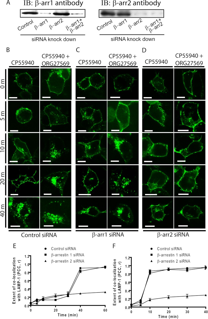
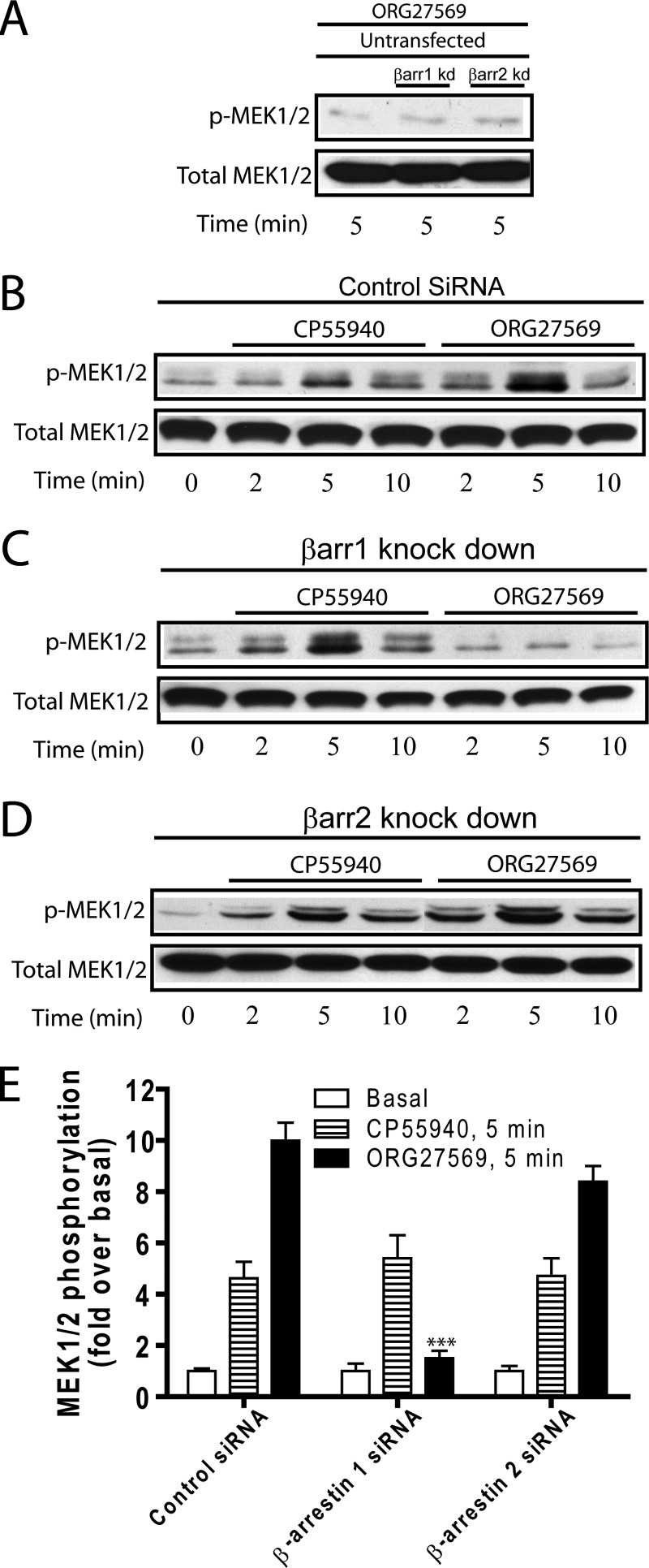
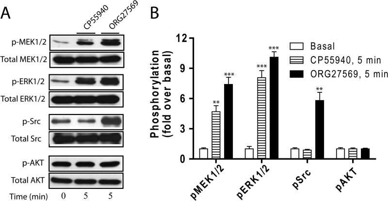
The cannabinoid receptor 1 (CB1) is a G protein-coupled receptor primarily expressed in brain tissue that has been implicated in several disease states. CB1 allosteric compounds, such as ORG27569, offer enormous potential as drugs over orthosteric ligands, but their mechanistic, structural, and downstream effects upon receptor binding have not been established. Previously, we showed that ORG27569 enhances agonist binding affinity to CB1 but inhibits G protein-dependent agonist signaling efficacy in HEK293 cells and rat brain expressing the CB1 receptor (Ahn, K. H., Mahmoud, M. M., and Kendall, D. A. (2012) J. Biol. Chem. 287, 12070-12082). Here, we identify the mediators of CB1 receptor internalization and ORG27569-induced G protein-independent signaling. Using siRNA technology, we elucidate an ORG27569-induced signaling mechanism for CB1 wherein β-arrestin 1 mediates short term signaling to ERK1/2 with a peak at 5 min and other upstream kinase components including MEK1/2 and c-Src. Consistent with these findings, we demonstrate co-localization of CB1-GFP with red fluorescent protein-β-arrestin 1 upon ORG27569 treatment using confocal microscopy. In contrast, we show the critical role of β-arrestin 2 in CB1 receptor internalization upon treatment with CP55940 (agonist) or treatment with ORG27569. These results demonstrate for the first time the involvement of β-arrestin in CB1-biased signaling by a CB1 allosteric modulator and also define the differential role of the two β-arrestin isoforms in CB1 signaling and internalization.


Figures

FIGURE 1.
FIGURE 1.
Effect of siRNA-mediated suppression of β-arrestin levels on cellular internalization of the CB1 T210A-GFP receptor in the absence or presence of ORG27569. A, the representative immunoblot (IB) depicts isoform-specific silencing of endogenous β-arrestin 1 (β-arr1) or β-arrestin 2 (β-arr2) expression by siRNAs. B–D, HEK293 cells were co-transfected with either control (B), β-arrestin 1 (C), or β-arrestin 2 (D) siRNAs and plasmid encoding the T210A-GFP receptor. The HEK293 cells were treated with 0.5 μm CP55940 in the presence of vehicle alone (0.03% Me2SO; left columns) or ORG27569 (10 μm; right columns) for the times indicated before fixation. Scale bars, 15 μm. E and F, the extent of co-localization of CB1 and LAMP-1 (the marker for the late endosome/lysosome) is quantified using the intensity correlation analysis for CP55940 treatment (n = 9) (E) and co-treatment of CP55940+ORG27569 (n = 9) (F). The PCC r was calculated at each time point (−1 = a negative correlation, 0 = no correlation, and +1 = a positive correlation). PCC (r) is presented as the means ± S.E.
FIGURE 2.
FIGURE 2.
Knockdown of β-arrestin 2 impairs the ORG27569-induced internalization of CB1 T210A-GFP. A–C, HEK293 cells were co-transfected with either control (A), β-arrestin 1 (B, β-arr1), or β-arrestin 2 (C, β-arr2) siRNAs and plasmid encoding the T210A-GFP receptor. The cells were incubated with 10 μm ORG27569 for 0, 3, and 4 h as indicated. After incubation, the cells were washed and fixed as described under “Experimental Procedures.” Localization of GFP-tagged receptor (green, left columns), the late endosome/lysosome marker, LAMP-1 (red, middle columns), and an overlay of the fluorescence images (yellow, right columns) are shown. The images are representative of at least three independent transfections that produced similar results. Scale bars, 15 μm (see A). D, the extent of co-localization quantified using the intensity correlation analysis is shown as described in the Fig. 1 legend. Nine images of each condition were analyzed (n = 9).
FIGURE 3.
FIGURE 3.
Inhibition of ORG27569-stimulated ERK1/2 phosphorylation by β-arrestin 1 siRNA using CB1 wild-type receptor. A, mock transfected HEK293 cells were used as control. B–D, HEK293 cells co-expressing CB1 wild type and either control (B), β-arrestin 1 (C, βarr1), or β-arrestin 2 (D, βarr2) siRNAs were exposed to CP55940 (0.5 μm) or ORG27569 (10 μm) for 0, 2, 5, or 10 min as indicated. Cell lysates were separated on SDS-PAGE and analyzed by Western blots probed with phospho-ERK1/2 (p-ERK1/2) followed by an HRP-conjugated anti-rabbit secondary antibody. The total level of ERK1/2 was detected for comparison. Immunoreactive signals were visualized by a chemiluminescent substrate system as described under “Experimental Procedures.” The levels of phosphorylation for the untransfected HEK293 cells are shown as a control. Representative blots of phosphorylated and total ERK1/2 of at least three separate experiments are shown for each condition. Note that the two bands correspond to the predominant isoforms, p42 (ERK2) and p44 (ERK1), for ERK signaling. E, graphs provide the quantified ERK1/2 phosphorylation levels for 5 and 10 min deduced from at least three experiments. The data are expressed as fold increases above the basal level of phosphorylation. The data represent the means ± S.E. of at least three independent experiments. Each treatment under the β-arrestin knockdown conditions was compared with its corresponding treatment under the control siRNA condition to determine the statistical significance of the differences using one-way analysis of variance and Bonferroni's post hoc test. *, p < 0.05; **, p < 0.01; ***, p < 0.005. F, β-arrestin 1 recruitment in HEK293 cells expressing wild-type CB1 receptor. HEK293 cells co-expressing the wild-type CB1-GFP and red fluorescent protein-β-arrestin 1 were exposed to ORG27569 for 0, 5, 15, 30, and 60 min. Cellular distribution of CB1-GFP and red fluorescent protein-β-arrestin 1 and the extent of co-localization between the two proteins were determined by confocal fluorescence microscopy as described in the legends to Figs. 1 and 2.
FIGURE 4.
FIGURE 4.
Inhibition of ORG27569-stimulated MEK1/2 phosphorylation by β-arrestin 1 siRNA using CB1 wild type. A, mock transfected HEK293 cells were used as control. B–D, HEK293 cells co-transfected with CB1 wild type and either control (B), β-arrestin 1 (C, βarr1), or β-arrestin 2 (D, βarr2) siRNAs were exposed to CP55940 (0.5 μm) or ORG27569 (10 μm) for 0, 2, 5, or 10 min as indicated. The cell lysates were analyzed by Western blots probed with phospho-MEK1/2 (p-MEK1/2) followed by an HRP-conjugated anti-rabbit secondary antibody. The total level of MEK1/2 was also detected for comparison using MEK1/2 antibody. The levels of phosphorylation for the mock transfected HEK293 cells are shown as a control. Representative blots of phosphorylated and total MEK1/2 of at least three separate experiments that produced similar results are shown for each condition. E, graphs provide the quantified MEK1/2 phosphorylation levels for 5 and 10 min deduced from at least three experiments. The data are expressed as fold increases above the basal level of phosphorylation. The data represent the means ± S.E. of at least three independent experiments. Each treatment under the β-arrestin knockdown conditions was compared with its corresponding treatment under the control siRNA condition to determine the statistical significance of the difference using one-way analysis of variance and Bonferroni's post hoc test. *, p < 0.05; **, p < 0.01; ***, p < 0.005.
FIGURE 5.
FIGURE 5.
Effect of siRNA-mediated suppression of β-arrestin levels on Src phosphorylation using CB1 wild type. A–C, HEK293 cells co-transfected with CB1 wild type and either control (A), β-arrestin 1 (B, βarr1), or β-arrestin 2 (C, βarr2) siRNAs were treated with CP55940 (0.5 μm) or ORG27569 (10 μm) for 0, 2, 5, or 10 min as indicated. The cell lysates were analyzed as described in Fig. 3 using phospho-Src and total Src antibodies. Representative blots of phosphorylated and total kinases of at least three separate experiments are shown for each condition. D, graphs provide the quantified phosphorylation levels for 5 and 10 min deduced from at least three experiments. The data are expressed as fold increases above the basal level of phosphorylation. The data represent the means ± S.E. of at least three independent experiments. Each treatment under the β-arrestin knockdown conditions was compared with its corresponding treatment under the control siRNA condition to determine the statistical significance of the differences using one-way analysis of variance and Bonferroni's post hoc test. *, p < 0.05; **, p < 0.01; ***, p < 0.005.
FIGURE 6.
FIGURE 6.
CP55940- and ORG27569-induced ERK1/2, MEK1/2, Src, and Akt signaling in hippocampal neuronal cells. A, rat hippocampal neurons endogenously expressing CB1 were exposed to CP55940 (0.5 μm) or ORG27569 (10 μm) for 5 min. Cell lysates were resolved by SDS-PAGE, and ERK1/2, MEK1/2, Src, and Akt phosphorylation was detected as described in the legend to Fig. 4. Representative blots of phosphorylated and total kinases are depicted. B, graphs show the quantified phosphorylation level of each kinase deduced from three experiments. The data are expressed as fold increases above basal levels of phosphorylation. The data represent the means ± S.E. of at least three independent experiments. Statistical significance of the differences was assessed using one-way analysis of variance and Bonferroni's post hoc test., *, p < 0.05; **, p < 0.01; ***, p < 0.005.
FIGURE 7.
FIGURE 7.
Proposed model of the ORG27569-induced β-arrestin 1-mediated ERK1/2 signaling. ORG27569 binding to CB1 promotes β-arrestin 1 recruitment which in turn recruits Src, MEK1/2, and ERK1/2 kinases to form a signaling complex.

References

    1. Price M. R., Baillie G. L., Thomas A., Stevenson L. A., Easson M., Goodwin R., McLean A., McIntosh L., Goodwin G., Walker G., Westwood P., Marrs J., Thomson F., Cowley P., Christopoulos A., Pertwee R. G., Ross R. A. (2005) Allosteric modulation of the cannabinoid CB1 receptor. Mol. Pharmacol. 68, 1484–1495 - PubMed
    1. Horswill J. G., Bali U., Shaaban S., Keily J. F., Jeevaratnam P., Babbs A. J., Reynet C., Wong Kai In P. (2007) PSNCBAM-1, a novel allosteric antagonist at cannabinoid CB1 receptors with hypophagic effects in rats. Br. J. Pharmacol. 152, 805–814 - PMC - PubMed
    1. Navarro H. A., Howard J. L., Pollard G. T., Carroll F. I. (2009) Positive allosteric modulation of the human cannabinoid (CB) receptor by RTI-371, a selective inhibitor of the dopamine transporter. Br. J. Pharmacol. 156, 1178–1184 - PMC - PubMed
    1. Zhang Y., Rodriguez A. L., Conn P. J. (2005) Allosteric potentiators of metabotropic glutamate receptor subtype 5 have differential effects on different signaling pathways in cortical astrocytes. J. Pharmacol. Exp. Ther. 315, 1212–1219 - PubMed
    1. Knudsen L. B., Kiel D., Teng M., Behrens C., Bhumralkar D., Kodra J. T., Holst J. J., Jeppesen C. B., Johnson M. D., de Jong J. C., Jorgensen A. S., Kercher T., Kostrowicki J., Madsen P., Olesen P. H., Petersen J. S., Poulsen F., Sidelmann U. G., Sturis J., Truesdale L., May J., Lau J. (2007) Small-molecule agonists for the glucagon-like peptide 1 receptor. Proc. Natl. Acad. Sci. U.S.A. 104, 937–942 - PMC - PubMed

Publication types

MeSH terms

LinkOut - more resources