. 2013 Apr 12;288(15):10684-91.
doi: 10.1074/jbc.M112.420042. Epub 2013 Feb 28.

Medium-chain fatty acid-sensing receptor, GPR84, is a proinflammatory receptor

Affiliations

Medium-chain fatty acid-sensing receptor, GPR84, is a proinflammatory receptor

Masakatsu Suzuki et al. J Biol Chem. .

Abstract

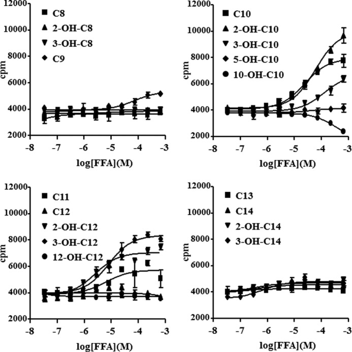
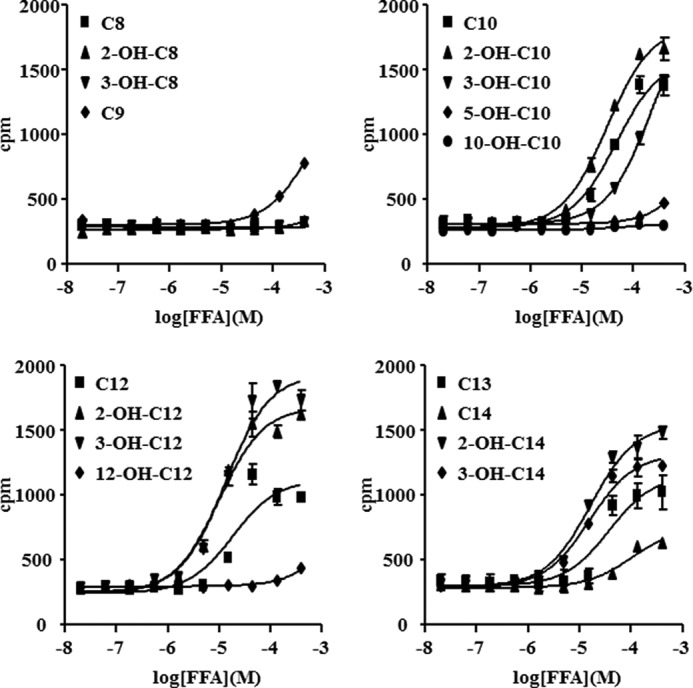
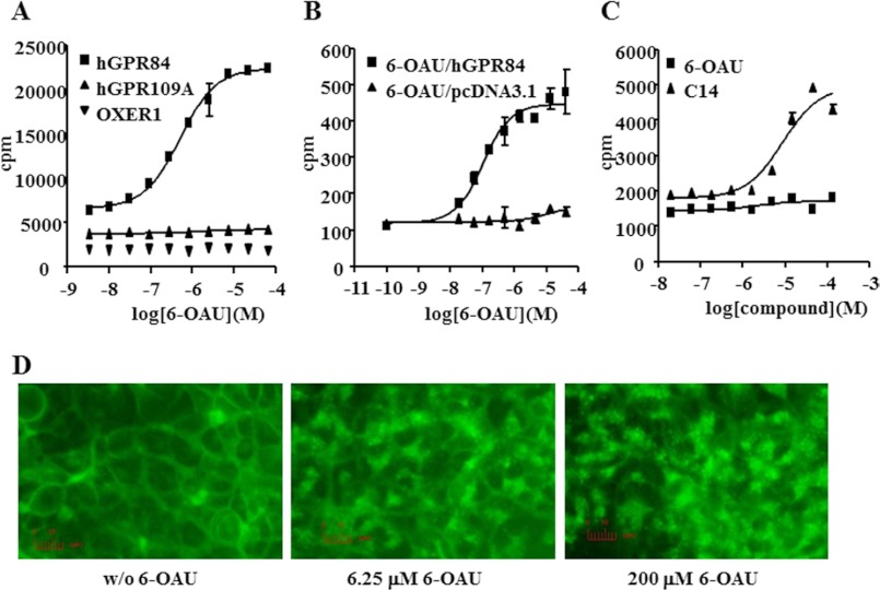
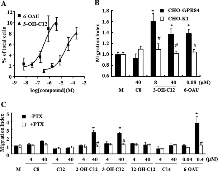
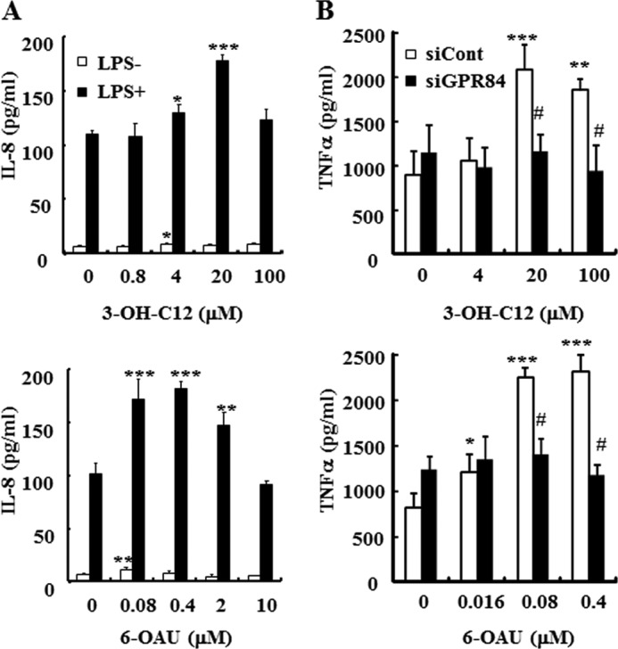
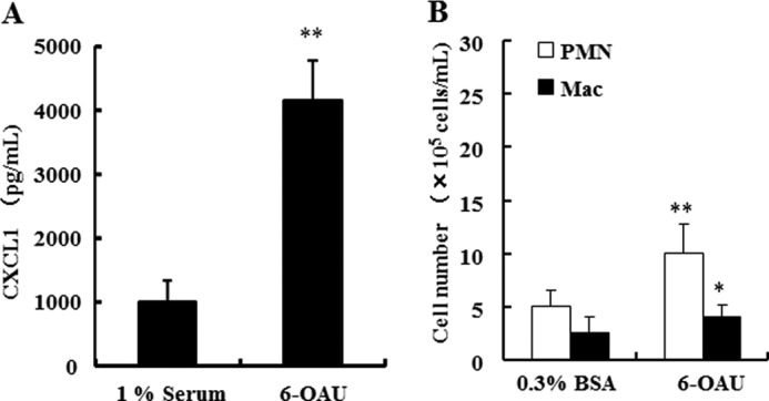
G protein-coupled receptor 84 (GPR84) is a putative receptor for medium-chain fatty acids (MCFAs), whose pathophysiological roles have not yet been clarified. Here, we show that GPR84 was activated by MCFAs with the hydroxyl group at the 2- or 3-position more effectively than nonhydroxylated MCFAs. We also identified a surrogate agonist, 6-n-octylaminouracil (6-OAU), for GPR84. These potential ligands and the surrogate agonist, 6-OAU, stimulated [(35)S]GTP binding and accumulated phosphoinositides in a GPR84-dependent manner. The surrogate agonist, 6-OAU, internalized GPR84-EGFP from the cell surface. Both the potential ligands and 6-OAU elicited chemotaxis of human polymorphonuclear leukocytes (PMNs) and macrophages and amplified LPS-stimulated production of the proinflammatory cytokine IL-8 from PMNs and TNFα from macrophages. Furthermore, the intravenous injection of 6-OAU raised the blood CXCL1 level in rats, and the inoculation of 6-OAU into the rat air pouch accumulated PMNs and macrophages in the site. Our results indicate a proinflammatory role of GPR84, suggesting that the receptor may be a novel target to treat chronic low grade inflammation associated-disease.


Figures

FIGURE 1.
FIGURE 1.
Potential ligands for GPR84. Membranes from Sf9 cells expressing human GPR84-fused with bovine Gαi protein were incubated with various FFAs for 1 h at room temperature. The data represent the means ± S.D. for quadruple determinations.
FIGURE 2.
FIGURE 2.
Effect of FFAs in PI assay. HEK293 cells were transfected with human GPR84 or control vector, pcDNA3.1, in the presence of Gqi5 plasmid. Cells were incubated with FFAs for 2 h at 37 °C. The data represent the means ± S.D. for triplicate determinations.
FIGURE 3.
FIGURE 3.
Identification and characterization of a surrogate agonist for GPR84. A, effect of 6-OAU on the [35S]GTPγS binding assay. Membranes from Sf9 cells expressing human GPR84 (hGPR84), human GPR109A (hGPR109A), or OXER1 fused with bovine Gαi protein were incubated with 6-OAU for 1 h at room temperature. The data represent the means ± S.D. for quadruple determinations. B, effect of 6-OAU on GPR84 in the PI assay. HEK293 cells were transfected with human GPR84 or control vector, pcDNA3.1, in the presence of Gqi5 plasmid. Cells were incubated with various concentrations of 6-OAU or 3-OH-C12 for 2 h at 37 °C. The data represent the means ± S.D. for triplicate determinations. C, effect of 6-OAU on GPR40 in the PI assay. HEK293 cells were transfected with human GPR40 or control vector, pcDNA3.1. Cells were incubated with various concentrations of 6-OAU or C14 for 2 h at 37 °C. The data represent the means ± S.D. for triplicate determinations. D, receptor internalization assay. Internalization of stably expressing human GPR84-EGFP fusion protein in HEK293 cells was observed by fluorescence microscopy. Cells were treated without or with 0, 6.25, or 200 μm 6-OAU for 30 min.
FIGURE 4.
FIGURE 4.
Cellular functions of human GPR84. A, chemotaxis of human peripheral PMNs. PMNs isolated from human peripheral blood were applied onto the upper chamber of a Transwell permeable support. Various concentrations of 6-OAU or 3-OH-C12 were added in the lower chambers of the Transwell. Transwell permeable supports were incubated at 37 °C for 1 h. The chemotaxis activities were assessed as a percentage of migrated cells to a lower chamber across the membrane from an upper chamber. The data were expressed as the means ± S.D. of quadruple determinations. B, chemotaxis of human GPR84 stable transfectant (CHO-GPR84). CHO-GPR84 was generated as described under “Experimental Procedures.” CHO-GPR84 cells were applied onto the upper chambers of the 96-well chemotaxis chamber. Various concentrations of 6-OAU or FFAs were added into the lower chambers. The chemotaxis activities were expressed as a migration index: (A595 of experimental well − A595 of background well)/(A595 of medium control well − A595 of background well). The data were expressed as means ± S.D.; n = 5. C, chemotaxis of U937 cell lines. U937 cells were used for the experiment after differentiation into macrophage-like cells as described under “Experimental Procedures.” Differentiated U937 cells were applied onto the upper chambers of the 96-well chemotaxis chamber. Various concentrations of 6-OAU or FFAs were added into the lower chambers. The chemotaxis activities were expressed as the same as B. The data were expressed as the means ± S.D. of triplicate determinations. *, p < 0.001 when compared with the medium control group (M). #p < 0.001 when compared with the PTX-untreated group.
FIGURE 5.
FIGURE 5.
Effects of 6-OAU or 3-OH-C12 on cytokine production. A, cytokine production from human peripheral PMNs. PMNs isolated from human peripheral blood were stimulated by LPS in the presence or absence of 6-OAU or 3-OH-C12. Culture supernatant was collected 4 h after stimulation. IL-8 concentration in the supernatant was measured with a commercial ELISA kit. The data were expressed as the means ± S.D. of triplicate determinations. *, p < 0.05, **, p < 0.01, and ***, p < 0.001 when compared with the agonist untreated group. B, negative control siRNA or GPR84 siRNA-transfected U937 macrophages were stimulated by 100 ng/ml LPS in the presence or absence of 6-OAU or 3-OH-C12. Culture supernatant was collected 16 h after stimulation. TNFα concentration in the supernatant was measured with a commercial ELISA kit. The data were expressed as the means ± S.D. of triplicate determinations. *, p < 0.05, **, p < 0.01, and ***, p < 0.001 when compared with the agonist untreated group. #, p < 0.01 when compared with the negative control siRNA-transfected group.
FIGURE 6.
FIGURE 6.
Proinflammatory effects of 6-OAU in vivo. A, 6-OAU (10 mg/kg) suspended in 1% rat serum or 1% rat serum as control was injected into the jugular vein in rats. Blood was collected from the jugular vein 3 h after 6-OAU injection. CXCL1 concentration in the serum was measured with a commercial ELISA kit. The data were expressed as the means ± S.D.; n = 5. **, p < 0.01 when compared with the 1% rat serum-injection group. B, 6-OAU suspended in 0.3% BSA or 0.3% BSA alone as control was injected into the dorsal air pouch prepared on rats. Infiltrated cells into the air pouch were collected with PBS 4 h after 6-OAU inoculation. The cell number was counted with the XT-2000i. The data were expressed as the means ± S.D.; n = 6. **, p < 0.01 and *, p < 0.05 when compared with the 0.3% BSA injection group.

References

    1. Wellendorph P., Johansen L. D., Bräuner-Osborne H. (2009) Molecular pharmacology of promiscuous seven transmembrane receptors sensing organic nutrients. Mol. Pharmacol. 76, 453–465 - PubMed
    1. Blad C. C., Tang C., Offermanns S. (2012) G protein-coupled receptors for energy metabolites as new therapeutic targets. Nat. Rev. Drug Discov. 11, 603–619 - PubMed
    1. Briscoe C. P., Tadayyon M., Andrews J. L., Benson W. G., Chambers J. K., Eilert M. M., Ellis C., Elshourbagy N. A., Goetz A. S., Minnick D. T., Murdock P. R., Sauls H. R., Jr., Shabon U., Spinage L. D., Strum J. C., Szekeres P. G., Tan K. B., Way J. M., Ignar D. M., Wilson S., Muir A. I. (2003) The orphan G protein-coupled receptor GPR40 is activated by medium and long chain fatty acids. J. Biol. Chem. 278, 11303–11311 - PubMed
    1. Itoh Y., Kawamata Y., Harada M., Kobayashi M., Fujii R., Fukusumi S., Ogi K., Hosoya M., Tanaka Y., Uejima H., Tanaka H., Maruyama M., Satoh R., Okubo S., Kizawa H., Komatsu H., Matsumura F., Noguchi Y., Shinohara T., Hinuma S., Fujisawa Y., Fujino M. (2003) Free fatty acids regulate insulin secretion from pancreatic beta cells through GPR40. Nature 422, 173–176 - PubMed
    1. Overton H. A., Babbs A. J., Doel S. M., Fyfe M. C., Gardner L. S., Griffin G., Jackson H. C., Procter M. J., Rasamison C. M., Tang-Christensen M., Widdowson P. S., Williams G. M., Reynet C. (2006) Deorphanization of a G protein-coupled receptor for oleoylethanolamide and its use in the discovery of small-molecule hypophagic agents. Cell Metab. 3, 167–175 - PubMed

MeSH terms