. 2013 Mar 15;74(1):1-29.
doi: 10.3114/sim0006. Epub 2012 Sep 14.

Development in Aspergillus

Affiliations

Development in Aspergillus

P Krijgsheld et al. Stud Mycol. .

Abstract

The genus Aspergillus represents a diverse group of fungi that are among the most abundant fungi in the world. Germination of a spore can lead to a vegetative mycelium that colonizes a substrate. The hyphae within the mycelium are highly heterogeneous with respect to gene expression, growth, and secretion. Aspergilli can reproduce both asexually and sexually. To this end, conidiophores and ascocarps are produced that form conidia and ascospores, respectively. This review describes the molecular mechanisms underlying growth and development of Aspergillus.

Keywords: Aspergillus; ascocarp; ascospore; asexual reproduction; conidiophore; conidium; development; fruiting body; fungi; heterogeneity; sexual reproduction; vegetative mycelium.


Figures

Fig. 1.
Fig. 1.
Scanning electron microscopy of cross sections of a 7 d old sandwiched A. niger colony. Cross sections were made at the periphery (A, D), four millimeter behind the periphery (B, E) and at the innermost center (C, F). The thickness of the colony is indicated by the distance between the white triangles. Panels D–F represent higher magnifications of A–C, respectively. Thin and thick arrows point at thin and thick hyphae, respectively. In H asterisks mark a non-sporulating conidiophore. Bars in panel C, for A–C, and F, for D–F, represent 100 and 20 μm.
Fig. 2.
Fig. 2.
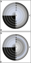
Growth (A, D), protein synthesis (B, E) and protein secretion (C, F) in a 7 d old xylose grown sandwiched colony of A. niger before (A–C) and after transfer (D–F) to fresh medium. (Adapted from Levin et al. 2007).
Fig. 3.
Fig. 3.
Development of A. niger monitored by scanning electron microscopy. The vegetative mycelium forms two types of aerial hyphae. One type is similar to vegetative hyphae (A), while the other type is 2–3 times thicker (B). The tips of the latter aerial hyphae may swell to form a vesicle (C,D). Buds are formed on the vesicle (E) that develop into metulae (F, G). Phialides are formed on top of the metulae (H), which give rise to chains of conidia (I, J). The bar in G also holds for A–F.
Fig. 4.
Fig. 4.
The central regulatory network consisting of BrlA, AbaA and WetA initiates asexual development in A. nidulans. StuA and MedA (A) and VosA (B) are regulators of brlA, abaA, and wetA.
Fig. 5.
Fig. 5.
Signaling cascades resulting in vegetative growth or asexual reproduction in A. nidulans. Signalling involves FluG (see Figure 6) and independently, two heterotrimeric G-protein complexes, both consisting of SfaD and GpgA (the Gβγ subunits) and the Gα subunits FadA and GanB, respectively. GTP-bound FadA and GanB stimulate vegetative growth via the cAMP PkaA pathway and repress asexual reproduction via brlA. The RGS proteins FlbA and RgsA hydrolyze the GTP bound to FadA and GanB, respectively, thereby repressing vegetative growth and promoting asexual development. The SfaD-GpgA dimer also stimulates vegetative growth. This is regulated by PhnA. (Adapted from Yu 2006).
Fig. 6.
Fig. 6.
Model of upstream regulation of brlA. FluG is involved in the formation of an extracellular factor that activates an unknown receptor. At a certain concentration of FluG, the general suppressor SfgA is inhibited removing the repression of the flb genes. FlbB and FlbE form a complex that activates brlA leading to asexual development. FlbC activates brlA together with the FlbB/FlbD transcription complex. FlbC also activates fluG and regulatory genes that act downstream of BrlA. FlbA activates brlA by inactivating FadA and probably plays a role in repressing fluG. (Adapted from Etxebeste et al. 2010).
Fig. 7.
Fig. 7.
GprA/B mediated signalling resulting in sexual development of A. nidulans. Signalling involves the heterotrimeric G-protein consisting of SfaD, GpgA and FadA. FadA bound to GTP activates a MAP kinase cascade (hypothetical protein AN2067.2, SteC, hypothetical protein AN3422.2 and MpkB). This in turn, activates the regulators NsdD and SteA that induce sexual development. In addition, SteA inhibits MedA that is also involved in activating sexual development. StuA, NsdC, and NosA also activate the sexual development program, while FlbC and FlbE are repressors of this pathway. RosA is a transcriptional inhibitor of veA, nsdD, nosA and stuA. (Adapted from Seo et al. 2004, Yu 2006).
Fig. 8.
Fig. 8.
Oxylipins, known as psi factor, regulate timing and balance between sexual and asexual development. The hormone like structures psiBα and psiCα stimulate sexual development, whereas psiAα and psiBβ stimulate asexual development. Psi factor results from ppoA, ppoB, and ppoC activity. Expression of these genes is regulated by the products resulting from the Ppo proteins and by BrlA and NsdD. In turn, the products resulting from the Ppo genes regulate expression of brlA and nsdD.
Fig. 9.
Fig. 9.
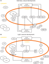
Light-regulated development in A. nidulans. (A) In the dark VelB enters the nucleus together with VeA and α-importin KapA. In the nucleus, VeA and VelB act as a dimeric complex or as a trimeric complex together with LaeA to positively regulate sexual development. VelB also forms a complex with VosA that negatively regulates asexual development. (B) In the light, activity of LaeA results in reduced levels of VelB and VosA. As a consequence, the inhibition of asexual development by the VelB-VosA complex is released. Moreover, the reduction of VelB levels abolishes stimulation of sexual development. Light is detected by the red light receptor FphA and the blue light receptor proteins LreA and LreB. These light receptors form a complex in the nucleus together with VeA. The cryptochrome/photolyase CryA also plays a role in light regulated development. Like FphA, it is a repressor of sexual development. (Adapted from Sarikaya Bayram et al. 2010).
Fig. 10.
Fig. 10.
Synthesis of melanin by means of the DHN pathway. Proteins of A. fumigatus responsible for each of the steps are indicated. Note that absence of particular enzymes and/or modification of melanin precursors will result in melanin-like pigments with colours other than brown/black. (Adapted from Fujii et al. 2004, Tsai et al. 1999, Pihet et al. 2009).
Fig. 11.
Fig. 11.
The cAMP/protein kinase A signaling pathway involved in germination of spores in A. nidulans. The presence of a carbon source is sensed by a GPCR that activates the Gα subunit GanB. GanB-GTP activates adenylate cyclase CyaA that produces cyclic adenosine-monophosphate (cAMP). cAMP binds to the regulatory subunit of PKA (PKAR), thus releasing the catalytic subunit PkaA. Active PkaA phosphorylates downstream targets resulting in swelling, germ tube formation and trehalose degradation. PkaA and PkaB have an overlapping role in spore germination in the presence of glucose but an opposite role in germination in the absence of a carbon source.

References

    1. Adams TH, Boylan MT, Timberlake WE. (1988). brlA is necessary and sufficient to direct conidiophore development in Aspergillus nidulans. Cell 54: 353–362 - PubMed
    1. Adams TH, Wieser JK, Yu JH. (1998). Asexual sporulation in Aspergillus nidulans. Microbiology and Molecular Biology Reviews 62: 35–54 - PMC - PubMed
    1. Agnihotri VP. (1964). Studies on Aspergilli. XVI. Effect of pH, temperature, and carbon and nitrogen interaction. Mycopathologia et Mycologia Applicata 24: 305–314 - PubMed
    1. Aguirre J. (1993). Spatial and temporal controls of the Aspergillus brlA developmental regulatory gene. Molecular Microbiology 8: 211–8 - PubMed
    1. Aimanianda V, Bayry J, Bozza S, Kniemeyer O, Perruccio K, et al. (2009). Surface hydrophobin prevents immune recognition of airborne fungal spores. Nature 460: 1117–1121 - PubMed