. 2013 Feb 28:4:31.
doi: 10.3389/fphys.2013.00031. eCollection 2013.

How cholesterol interacts with membrane proteins: an exploration of cholesterol-binding sites including CRAC, CARC, and tilted domains

Affiliations

How cholesterol interacts with membrane proteins: an exploration of cholesterol-binding sites including CRAC, CARC, and tilted domains

Jacques Fantini et al. Front Physiol. .

Abstract

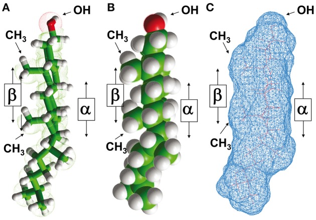
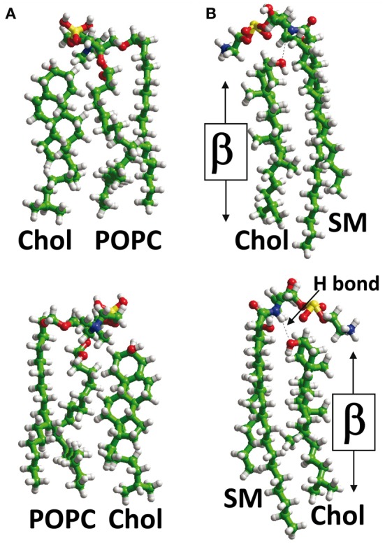
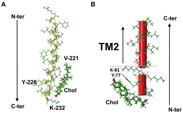
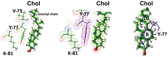
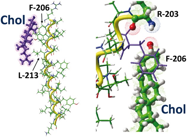
The plasma membrane of eukaryotic cells contains several types of lipids displaying high biochemical variability in both their apolar moiety (e.g., the acyl chain of glycerolipids) and their polar head (e.g., the sugar structure of glycosphingolipids). Among these lipids, cholesterol is unique because its biochemical variability is almost exclusively restricted to the oxidation of its polar -OH group. Although generally considered the most rigid membrane lipid, cholesterol can adopt a broad range of conformations due to the flexibility of its isooctyl chain linked to the polycyclic sterane backbone. Moreover, cholesterol is an asymmetric molecule displaying a planar α face and a rough β face. Overall, these structural features open up a number of possible interactions between cholesterol and membrane lipids and proteins, consistent with the prominent regulatory functions that this unique lipid exerts on membrane components. The aim of this review is to describe how cholesterol interacts with membrane lipids and proteins at the molecular/atomic scale, with special emphasis on transmembrane domains of proteins containing either the consensus cholesterol-binding motifs CRAC and CARC or a tilted peptide. Despite their broad structural diversity, all these domains bind cholesterol through common molecular mechanisms, leading to the identification of a subset of amino acid residues that are overrepresented in both linear and three-dimensional membrane cholesterol-binding sites.

Keywords: Alzheimer; CH-Pi; alpha-synuclein; cholesterol; lipid raft; lipid-protein interaction; neurotransmitter; receptor structure.


Figures

Figure 1
Figure 1
Structural properties of cholesterol. The asymmetric distribution of aliphatic groups (methyl, iso-octyl) linked to the planar sterane backbone of cholesterol defines two distinct sides referred to as α and β faces, according to the nomenclature of ring compounds proposed by Rose et al. (1980). This asymmetric structure of cholesterol is illustrated in a tube model (A), a sphere model (B), and a molecular surface model (C). Note that the OH group is closer to the “smooth” α face than to the “rough” β face.
Figure 2
Figure 2
Lipid-cholesterol interactions. In the plasma membrane, cholesterol (Chol) can interact with phosphatidylcholine, e.g., palmitoyl-oleyl-phosphatidylcholine (POPC) (panel A) or sphingolipids such as sphingomyelin (panel B). When cholesterol interacts with POPC, its OH group is not buried in the complex, and both its α and β faces are available for TM domains of proteins (A). However, when cholesterol interacts with SM, a hydrogen bond (H bond) is formed between the OH group of cholesterol and the NH group of the sphingolipid. This H bond orientates cholesterol with respect to SM so that only its β face remains available for TM domains. The OH group of cholesterol is masked by the polar head of sphingomyelin in a typical “umbrella” effect.
Figure 3
Figure 3
Cholesterol-cholesterol interactions. In model membranes, two cholesterol molecules can form a tail-to-tail (A) or a face-to-face (B) complex. In the latter case, the self-recognition properties of cholesterol can induce the dimerization of membrane receptors (C), as demonstrated for G-protein-coupled receptors with 7-TM domains.
Figure 4
Figure 4
The CRAC/cholesterol complex in a membrane environment. (A) Docking of cholesterol on the CRAC domain of the TM5 domain of human type 3 somatostatin receptor. The CRAC domain (221-VICLCYLLIVVKK-232) is located in the cytoplasmic leaflet of the membrane bilayer. Note that the central Y-226 residue of CRAC is not involved in cholesterol interaction. The total energy of interaction has been estimated at −43 kJ.mol−1 (Baier et al., 2011). (B) Docking of cholesterol on the CRAC domain close the TM2 domain of human delta-type opioid receptor. In this case, the CRAC domain (fragment 74-IVRYTKMK-81) lies outside the membrane. The aromatic side chain of Y-77 plays a critical role in this interaction (see Figure 5). The polar-apolar interface of the membrane is indicated by a dotted gray line.
Figure 5
Figure 5
Molecular mechanisms of cholesterol-CRAC interaction. This figure shows a detailed analysis of the interaction between cholesterol and the CRAC domain of the human delta-type opioid receptor (see Figure 4). Three distinct views of the complex are shown, with residues I-74, Y-77, and K-81 enlightened. The NH+3 of K-81, and the OH groups of Y-77 and cholesterol are rejected in a polar area where they can form a network of energetically favored electrostatic interactions (including hydrogen bonds). The aromatic side chain of Y-77 stacks onto the B ring of sterane backbone through typical CH-π stacking interactions. The isooctyl chain of cholesterol interacts with the aliphatic side chains of I-74 (not shown) and V-75. The sterane rings are indicated for cholesterol in the right panel.
Figure 6
Figure 6
The CARC/cholesterol complex. This figure shows the docking of cholesterol on the CARC domains of the TM5 domain of the human type 3 somatostatin receptor. The CARC domain (fragment 203-RAGFIIYTAAL-213) is located in the extracellular leaflet of the TM5 domain of the receptor. Two distinct views of the complex are shown, one with the whole TM5 domain (left panel), the other with residues R-203 and F-206 enlightened. Note the CH-π stacking interaction of the phenyl ring of F-206 onto the A ring of the sterane backbone of cholesterol. The large aliphatic chain of L-213 interacts with the isooctyl group of cholesterol.
Figure 7
Figure 7
Occurrence of two cholesterol binding motifs (CRAC and CARC) in the same TM domain. An example of the simultaneous occurrence of two cholesterol-recognition motifs in the same TM segment is given by the 5th TM domain of the human type 3 somatostatin receptor, which possesses a CRAC domain in the cytoplasmic leaflet (in blue) and a CARC domain in its exofacial leaflet (in yellow). The calculated energy of interaction of each domain with cholesterol is indicated. The surface rendering of the TM domain is particularly suited to visualize the three-dimensional interaction with cholesterol (rendered as space fill models).
Figure 8
Figure 8
How cholesterol interacts with a tilted peptide. Docking of cholesterol with the N-terminal part of HIV-1 gp41, i.e., the fusion peptide. The apolar part of cholesterol interacts with the aromatic ring of F-8 through CH-π (but not stacking) interactions. The OH group of cholesterol is close to the α-NH3+ group of the peptide (N-terminus ending). The angle between the helix axis of the tilted fragment of the fusogenic tilted peptide of gp41 is 41°. The calculated energy of interaction is −48.5 kJ.mol−1.

References

    1. Anderson R. G., Jacobson K. (2002). A role for lipid shells in targeting proteins to caveolae, rafts, and other lipid domains. Science 296, 1821–1825 10.1126/science.1068886 - DOI - PubMed
    1. Baier C. J., Fantini J., Barrantes F. J. (2011). Disclosure of cholesterol recognition motifs in transmembrane domains of the human nicoticin acetylcholine receptor. Sci. Reports 1:69 10.1038/srep00069 - DOI - PMC - PubMed
    1. Ballesteros J., Weinstein H. (1995). Integrated methods for the construction of three-dimensional models and computational probing of structure-function relations in G protein-coupled receptors. Methods Neurosci. 25, 366–428
    1. Barrantes F. J., Borroni V., Vallés S. (2010). Neuronal nicotinic acetylcholine receptor-cholesterol crosstalk in Alzheimer's disease. FEBS Lett. 584, 1856–1863 10.1016/j.febslet.2009.11.036 - DOI - PubMed
    1. Barrett P. J., Song Y., Van Horn W. D., Hustedt E. J., Schafer J. M., Hadziselimovic A., et al. (2012). The amyloid precursor protein has a flexible transmembrane domain and binds cholesterol. Science 336, 1168–1171 10.1126/science.1219988 - DOI - PMC - PubMed