. 2013;8(2):e56982.
doi: 10.1371/journal.pone.0056982. Epub 2013 Feb 22.

Antineoplastic effects of α-santalol on estrogen receptor-positive and estrogen receptor-negative breast cancer cells through cell cycle arrest at G2/M phase and induction of apoptosis

Affiliations

Antineoplastic effects of α-santalol on estrogen receptor-positive and estrogen receptor-negative breast cancer cells through cell cycle arrest at G2/M phase and induction of apoptosis

Sreevidya Santha et al. PLoS One. 2013.

Erratum in

  • PLoS One. 2013;8(6). doi:10.1371/annotation/c732480c-eb97-4eff-acb6-300797f4efa9

Abstract

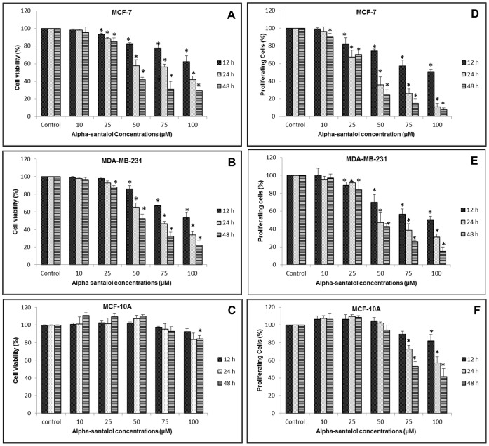
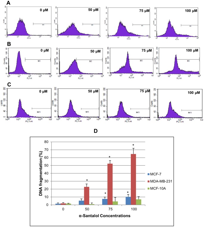
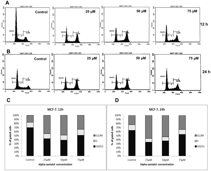
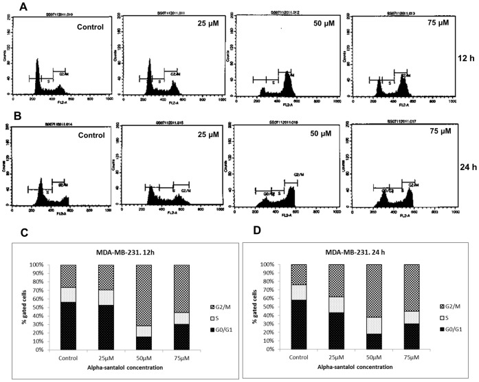
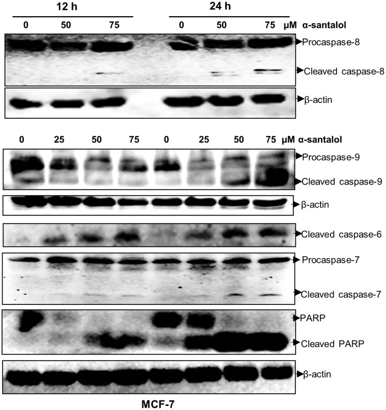
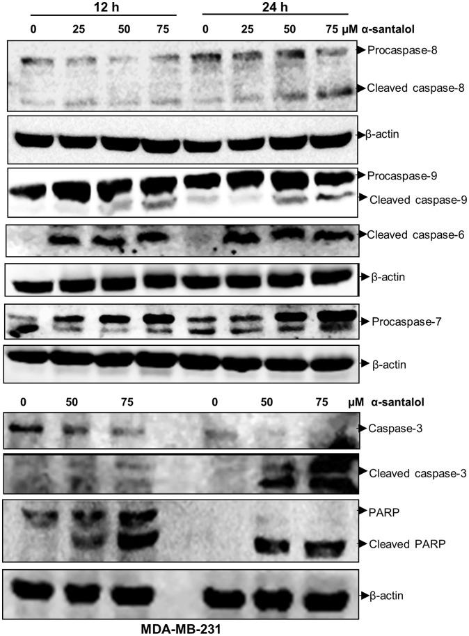
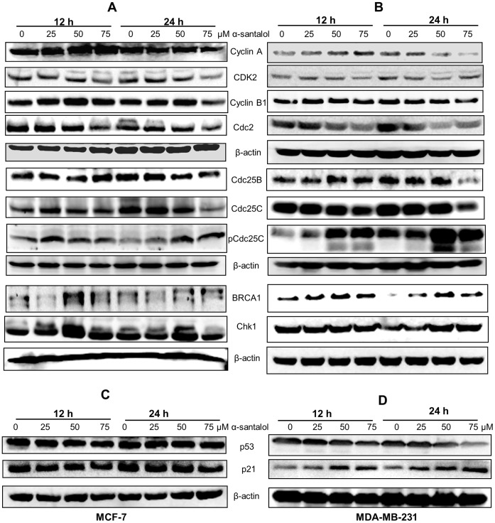
Anticancer efficacy and the mechanism of action of α-santalol, a terpenoid isolated from sandalwood oil, were investigated in human breast cancer cells by using p53 wild-type MCF-7 cells as a model for estrogen receptor (ER)-positive and p53 mutated MDA-MB-231 cells as a model for ER-negative breast cancer. α-Santalol inhibited cell viability and proliferation in a concentration and time-dependent manner in both cells regardless of their ER and/or p53 status. However, α-santalol produced relatively less toxic effect on normal breast epithelial cell line, MCF-10A. It induced G2/M cell cycle arrest and apoptosis in both MCF-7 and MDA-MB-231 cells. Cell cycle arrest induced by α-santalol was associated with changes in the protein levels of BRCA1, Chk1, G2/M regulatory cyclins, Cyclin dependent kinases (CDKs), Cell division cycle 25B (Cdc25B), Cdc25C and Ser-216 phosphorylation of Cdc25C. An up-regulated expression of CDK inhibitor p21 along with suppressed expression of mutated p53 was observed in MDA-MB-231 cells treated with α-santalol. On the contrary, α-santalol did not increase the expression of wild-type p53 and p21 in MCF-7 cells. In addition, α-santalol induced extrinsic and intrinsic pathways of apoptosis in both cells with activation of caspase-8 and caspase-9. It led to the activation of the executioner caspase-6 and caspase-7 in α-santalol-treated MCF-7 cells and caspase-3 and caspase-6 in MDA-MB-231 cells along with strong cleavage of poly(ADP-ribose) polymerase (PARP) in both cells. Taken together, this study for the first time identified strong anti-neoplastic effects of α-santalol against both ER-positive and ER-negative breast cancer cells.


Conflict of interest statement

Competing Interests: The authors have declared that no competing interests exist.

Figures

Figure 1
Figure 1. Effect of α-santalol on cell viability and proliferation.
Human breast cancer cells MCF-7 and MDA-MB-231 and normal human breast epithelial cells MCF-10A were treated with either DMSO (control) or 10–100 µM α-santalol for 12, 24 and 48 h. At the end of respective treatments, MTT and BrdU incorporation assays were performed on each cell line. Data in left panels (A–C) were obtained from MTT assays and data in right panels (D–F) were from BrdU cell proliferation ELISA of MCF-7, MDA-MB-231 and MCF-10A cells respectively. Values were shown as mean ± SD of at least three experiments. *, P<0.05 indicates statistically significant decrease in α-santalol treated groups as compared with the control.
Figure 2
Figure 2. Effect of α-santalol on DNA fragmentation by TUNEL assay and flow cytometry.
(A) MCF-7, (B) MDA-MB-231 and (C) MCF-10A cells were treated with α-santalol (0–100 µM) for 48 h and the extent of DNA fragmentation was determined by flow cytometric analysis. APO-BrdU TUNEL assay kit (Invitrogen) was used for the experiment and BrdU incorporation at DNA strand breaks of apoptotic cells were detected by conjugation to an Alexa Fluor 488 dye-labeled anti-BrdU antibody. The extent of DNA fragmentation was quantified by computational analysis of cells staining positive for BrdU using CellQuest software. (D) The bar graph indicates the percentages of apoptotic cells with fragmented DNA in MCF-7, MDA-MB-231 and MCF-10A cells. In each case data represents mean ± SD of three observations. *, P<0.05 indicates statistical significance in α-santalol treated groups compared with the control.
Figure 3
Figure 3. Effect of α-santalol on cell cycle progression in MCF-7 cells.
Cells were treated with DMSO (control) or 25–75 µM of α-sanatlol for 12 and 24 h, stained with propidium iodide and distribution of cells in different phases of cell cycle were analyzed by flow cytometer. Histograms representing the fluorescence pattern for cell cycle distribution in different treatments are shown for 12 h (A) and 24 h (B). The percentage of cells in each cell cycle phases after 12 h and 24 h respectively are shown in (C) and (D). Data shown here are representative of those having similar results.
Figure 4
Figure 4. Effect of α-santalol on cell cycle progression in MDA-MB-231 cells.
Data were obtained as explained in figure 3. Representative histograms of the fluorescence pattern for cell cycle distribution in different treatments after 12 h (A) and 24 h (B) are shown. (C) and (D) are the percentages of MDA-MB-231 cells in each cell cycle phases after 12 and 24 h α-santalol treatment.
Figure 5
Figure 5. α-Santalol induces activation of different caspases and PARP cleavage in MCF-7 cells.
Total cell lysates were prepared from the cells treated with the indicated concentration of α-sanatlol for 12 and 24 h. Equal amounts of proteins were separated by SDS-PAGE and subjected to western immunoblotting. Membranes were probed with respective primary antibodies followed by appropriate secondary antibody and protein expression was determined by ECL detection system. β-actin was used to verify equal loading of the samples.
Figure 6
Figure 6. α-Santalol induces apoptosis in MDA-MB-231 cells by activating different caspases and PARP cleavage.
Total cell lysates were made and the expressions of various apoptotic proteins were determined by western immunoblotting. β-actin was used as an internal control.
Figure 7
Figure 7. Western Blot analysis of the cell cycle regulatory proteins.
(A) MCF-7 and (B) MDA-MB-231 cells were treated with α-santalol (0–75 µM) for 12 and 24 h, and equal amounts of proteins were subjected to immunoblot for the detection of the indicated G2/M regulatory proteins. (C) and (D) show the effect of α-santalol on p21 and p53 expression in MCF-7 and MDA-MB-231 cells respectively. Blots were probed for β-actin to ensure equal protein loading.

Similar articles

Cited by

References

    1. Zhang X, Dwivedi C (2011) Skin cancer chemoprevention by α-santalol. Front Biosci (Schol Ed) 3: 777–787. - PubMed
    1. Burdock GA, Carabin IG (2008) Safety assessment of sandalwood oil (Santalum album L.) Food Chem Toxicol. 46: 421–432. - PubMed
    1. Dwivedi C, Guan X, Harmsen WL, Voss AL, Goetz-Parten DE, et al. (2003) Chemopreventive effects of α-santalol on skin tumor development in CD-1 and SENCAR mice. Cancer Epidemiol Biomarkers Prev 12: 151–156. - PubMed
    1. Dwivedi C, Valluri HB, Guan X, Agarwal R (2006) Chemopreventive effects of α-santalol on ultraviolet B radiation-induced skin tumor development in SKH-1 hairless mice. Carcinogenesis 27: 1917–1922. - PubMed
    1. Kaur M, Agarwal C, Singh RP, Dwivedi C, Agarwal R (2005) Skin cancer chemopreventive agent, α-santalol, induces apoptotic death of human epidermoid carcinoma A431 cells via caspase activation together with dissipation of mitochondrial membrane potential and cytochrome c release. Carcinogenesis 26: 369–380. - PubMed

Publication types