. 2013;8(2):e57128.
doi: 10.1371/journal.pone.0057128. Epub 2013 Feb 22.

Functional capacity of Shiga-toxin promoter sequences in eukaryotic cells

Affiliations

Functional capacity of Shiga-toxin promoter sequences in eukaryotic cells

Leticia V Bentancor et al. PLoS One. 2013.

Abstract

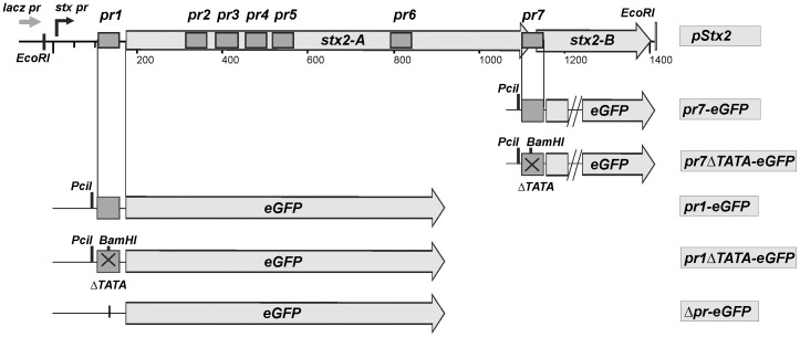
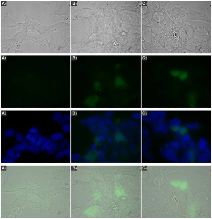
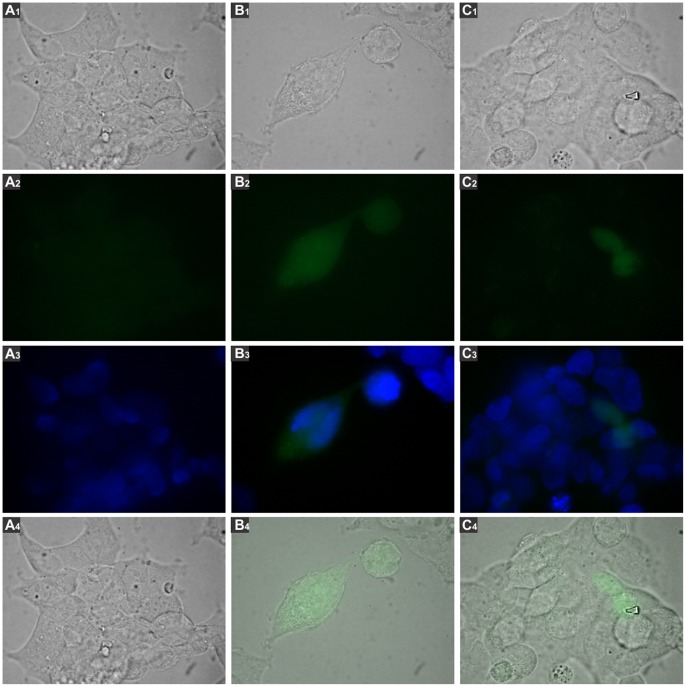
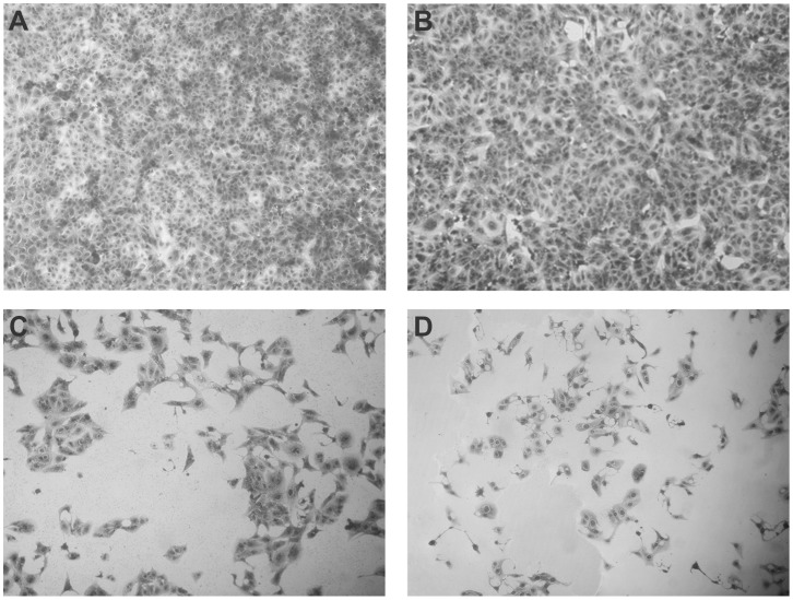
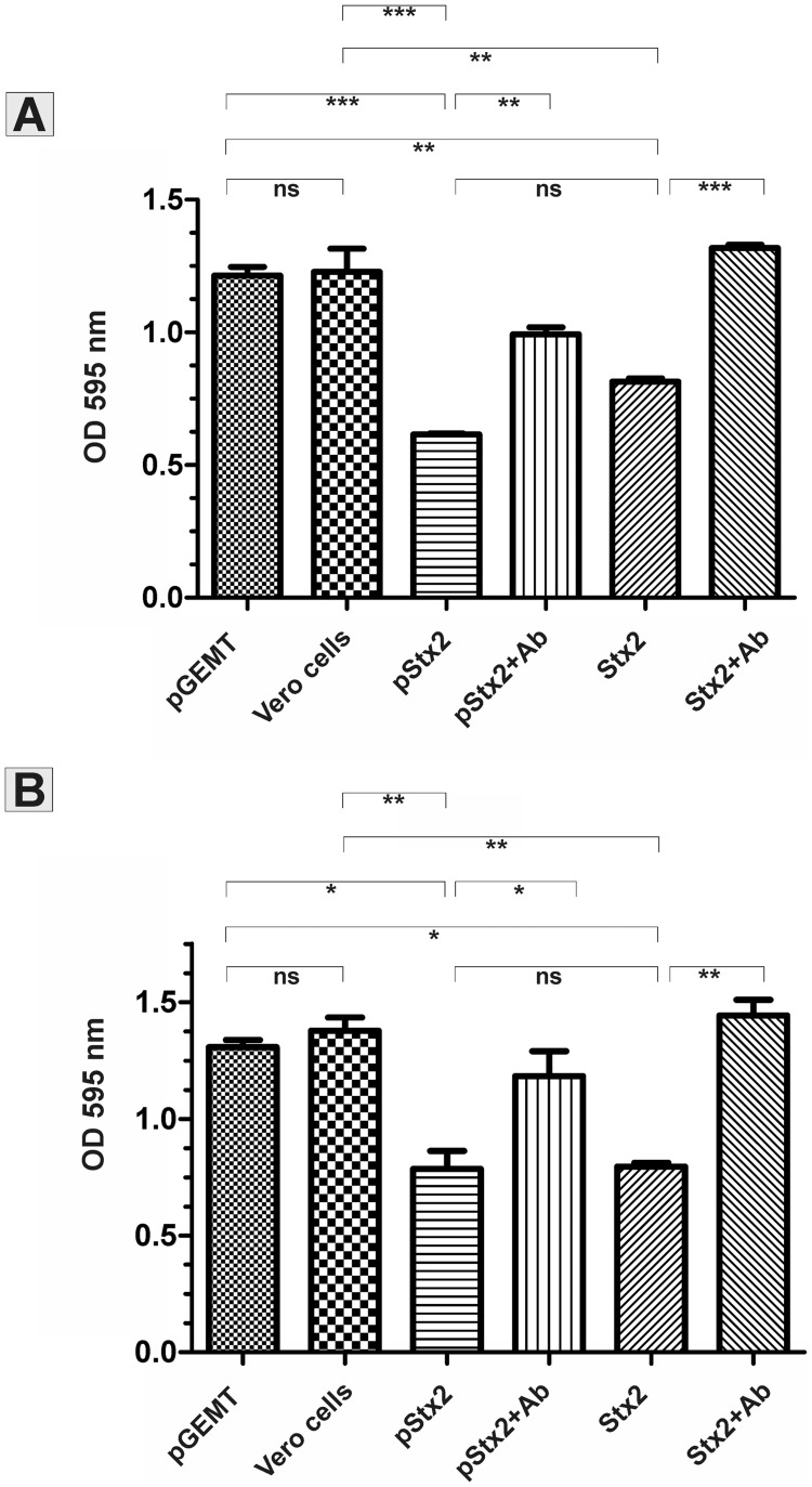
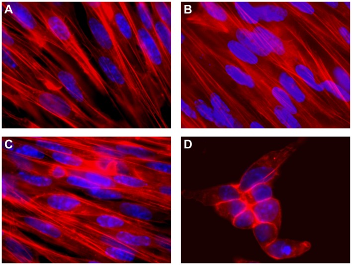
Shiga toxins (Stx) are the main virulence factors in enterohemorrhagic Escherichia coli (EHEC) infections, causing diarrhea and hemolytic uremic syndrome (HUS). The genes encoding for Shiga toxin-2 (Stx2) are located in a bacteriophage. The toxin is formed by a single A subunit and five B subunits, each of which has its own promoter sequence. We have previously reported the expression of the B subunit within the eukaryotic environment, probably driven by their own promoter. The aim of this work was to evaluate the ability of the eukaryotic machinery to recognize stx2 sequences as eukaryotic-like promoters. Vero cells were transfected with a plasmid encoding Stx2 under its own promoter. The cytotoxic effect on these cells was similar to that observed upon incubation with purified Stx2. In addition, we showed that Stx2 expression in Stx2-insensitive BHK eukaryotic cells induced drastic morphological and cytoskeletal changes. In order to directly evaluate the capacity of the wild promoter sequences of the A and B subunits to drive protein expression in mammalian cells, GFP was cloned under eukaryotic-like putative promoter sequences. GFP expression was observed in 293T cells transfected with these constructions. These results show a novel and alternative way to synthesize Stx2 that could contribute to the global understanding of EHEC infections with immediate impact on the development of treatments or vaccines against HUS.


Conflict of interest statement

Competing Interests: The authors have declared that no competing interests exist.

Figures

Figure 1
Figure 1. In silico analysis of the stx2 sequence.
A. Seven regions (pr1-7) with high score for putative eukaryotic promoter sequences were found. The putative transcription start site (TSS, +1) detected with promoter prediction server is highlighted in gray B. The pr1-pr7 regions are indicated with dark gray boxes over the stx2 gene. Putative mammalian transcription factor binding sites are indicated on the corresponding sequences of the pr1 and pr7 regions.
Figure 2
Figure 2. Recombinant plasmid constructs.
pStx2 is the pGEM-T plasmid with the stx2 locus cloned in the same direction that lacZ promoter. pr1-eGFP and pr7-eGFP are reporter plasmids with the region pr1 or pr7 driving eGFP expression, and Δpr-eGFP is a construction lacking the wild CMV promoter and was used as negative control. Linear pr1-eGFP and pr7-eGFP were obtained after digestion with PciI. In pr1ΔTATA-eGFP and pr7ΔTATA-eGFP TATA Box sequence has been replaced by BamHI restriction site. Stx2 linear DNA was obtained after digestion with EcoRI.
Figure 3
Figure 3. GFP activity driven by the pr1 or pr7 regions.
293 T cells were transfected with plasmids pr1-eGFP, pr7-eGFP or Δpr-eGFP, incubated for 48 h and analyzed by fluorescence microscopy using the Nikon Eclipse TE2000 microscope equipped with a CCD camera, with 1000X magnification. Green fluorescence photos were taken with 400 ms of exposure and 3.2 of gain. Numbers 1, 2, 3, 4 correspond to images visualized with white light, green filter, merge between DAPI and green filter and merge between white light and green filter, respectively. A. Cells transfected with the Δpr-eGFP plasmid. B. Cells transfected with pr1-eGFP. C. Cells transfected with pr7-eGFP.
Figure 4
Figure 4. GFP activity driven by linear reporter plasmid.
293 T cells were transfected with pr1-eGFP or pr7-eGFP linearized with Pcil restriction enzyme. After 48 h, cells were analyzed by fluorescence microscopy using Nikon Eclipse TE2000 microscope equipped with a CCD camera, using 1000X magnification. Green fluorescence photos were taken with 400 ms of exposure and 3.2 of gain. Numbers 1, 2, 3, 4 correspond to images visualized with white light, green filter, merge between DAPI and green filter and merge between white light and green filter, respectively. A. 293 T cells transfected with the Δpr-eGFP plasmid. B. Cells transfected with linear pr1-eGFP. C. Cells transfected with linear pr7-eGFP.
Figure 5
Figure 5. Immunofluorescence assay of Vero cells incubated with Stx2.
Cells were incubated with purified Stx2 (100 CD50) or transfected with the pStx2 plasmid and stained with propidium iodide. Presence of Stx2 was detected with polyclonal anti-Stx2 serum and FITC-conjugated goat anti-mouse antibody, and analyzed by confocal microscopy. Original magnification 600X. A: Non-treated Vero cells. B: Cells transfected with the pGEM-T vector alone. C: Cells incubated with purified Stx2. D: Digital zoom (3.5X) of picture C. E: Cells transfected with pStx2. F: Digital zoom (3.5X) of picture E.
Figure 6
Figure 6. Cytotoxicity on Vero Cells.
Vero cells were incubated with purified Stx2 (1 CD50) or transfected with pStx2 plasmid. After 48 h, cells were stained with Crystal Violet and analyzed by optical microscopy. Representative pictures using 200X original magnification are shown. A. Non-treated Vero cells. B. Cells transfected with the pGEM-T plasmid. C. Cells transfected with the pStx2 plasmid. D. Cells incubated with purified Stx2.
Figure 7
Figure 7. Neutralization of the Stx2 cytotoxic activity.
Vero cells were incubated with a 1∶1600 dilution of cellular extracts (Panel A) or a 1∶400 dilution of culture supernatants (Panel B) derived from Vero cells transfected with pGEM-T (pGEM-T) or pStx2 (pStx2). As positive and negative controls, Vero cells were incubated with 1 CD50 of Stx2 (Stx2) or in medium (Vero cells), respectively. To evaluate the specificity of the cytotoxicity, cytotoxic samples were pre-incubated with mouse polyclonal anti-Stx2 antibodies (pStx2+Ab; Stx2+Ab). After 48 h, cells were stained with Crystal Violet and OD595 was measured as detailed in Materials and Methods. One-way ANOVA (Tukey’s Multiple Comparison Test) was used to determine statistical significance between different samples.*P<0.05. **P<0.01. ***P<0.001.
Figure 8
Figure 8. Cytotoxicity on BHK cells.
BHK cells were incubated with purified Stx2 (1 CD50) or transfected with the pStx2 plasmid. After 48 h, cells were stained with DAPI and phalloidin-TRITC and analyzed by fluorescence microscopy. Original magnification 600X. A. Non-treated BHK cells. B. Cells transfected with pGEM-T. C. Cells incubated with purified Stx2. D. Cells transfected with the pStx2 plasmid.
Figure 9
Figure 9. Transduction of THP-1 cells differentiated to macrophages by bacteriophage 933W.
THP-1 cells PMA-differentiated to macrophages were transduced with φΔTOX-GFP. After 3 h, cells were analyzed by confocal microscopy, using 600X magnification. Green fluorescence photos were taken with 400 ms of exposure and 1 of gain. Numbers 1, 2, 3, 4 correspond to images visualized with white light, red filter, green filter and merge between green and red filter, respectively. A. Cells transduced with φΔTOX-GFP. B. Non-treated cells.

References

    1. Griffin PM, Tauxe RV (1991) The epidemiology of infections caused by Escherichia coli O157:H7, other enterohemorrhagic E. coli, and the associated hemolytic uremic syndrome. Epidemiol Rev 13: 60–98. - PubMed
    1. Slutsker L, Ries AA, Greene KD, Wells JG, Hutwagner L, et al. (1997) Escherichia coli O157:H7 diarrhea in the United States: clinical and epidemiologic features. Ann Intern Med 126: 505–513. - PubMed
    1. Caprioli A, Morabito S, Brugere H, Oswald E (2005) Enterohaemorrhagic Escherichia coli: emerging issues on virulence and modes of transmission. Vet Res 36: 289–311. - PubMed
    1. Lopez EL, Prado-Jimenez V, O’Ryan-Gallardo M, Contrini MM (2000) Shigella and Shiga toxin-producing Escherichia coli causing bloody diarrhea in Latin America. Infect Dis Clin North Am 14: 41–65. - PubMed
    1. Rivas M, Miliwebsky E, Chinen I, Deza N, Leotta GA (2006) The epidemiology of hemolytic uremic syndrome in Argentina. Diagnosis of the etiologic agent, reservoirs and routes of transmission. Medicina 66: 27–32. - PubMed

Publication types

LinkOut - more resources