. 2013;8(2):e57571.
doi: 10.1371/journal.pone.0057571. Epub 2013 Feb 25.

VPO1 mediates ApoE oxidation and impairs the clearance of plasma lipids

Affiliations

VPO1 mediates ApoE oxidation and impairs the clearance of plasma lipids

Youfeng Yang et al. PLoS One. 2013.

Abstract

Objective: ApoE is an abundant component of chylomicron, VLDL, IDL, and HDL. It binds to multiple types of lipids and is implicated in cholesterol and triglyceride homeostasis. Oxidation of ApoE plays a crucial role in the genesis of atherosclerosis. It is proposed that heme-containing peroxidases (hPx) are major mediators of lipoprotein oxidization. Vascular peroxidase 1 (VPO1) is a recently-discovered hPx, which is expressed in cardiovascular system, lung, liver etc. and secreted into plasma. Its plasma concentration is three orders of magnitude of that of myeloperoxidase. If VPO1 mediates ApoE oxidation and affects the lipid metabolism remains to be elucidated.

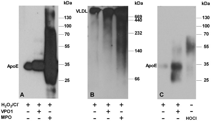
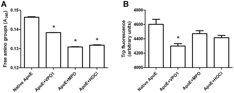
Methods: Recombinant ApoE and VPO1 were expressed and purified from stably-expressing cell lines deriving from HEK293 cells. ApoE oxidation was carried out by VPO1 in the presence of H2O2 and chloride. ApoE oxidation was verified by a variety of approaches including immunoblot and amino acid analyses. To evaluate the functional changes in VPO1-oxidized ApoE, lipid emulsion particle binding assays were employed.

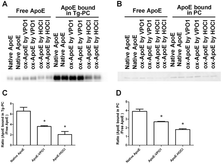
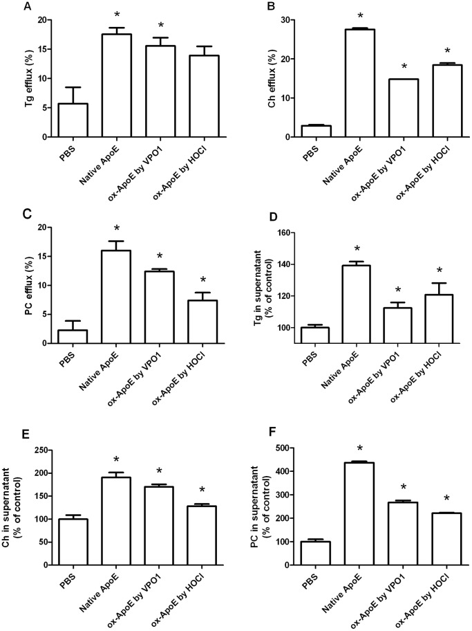
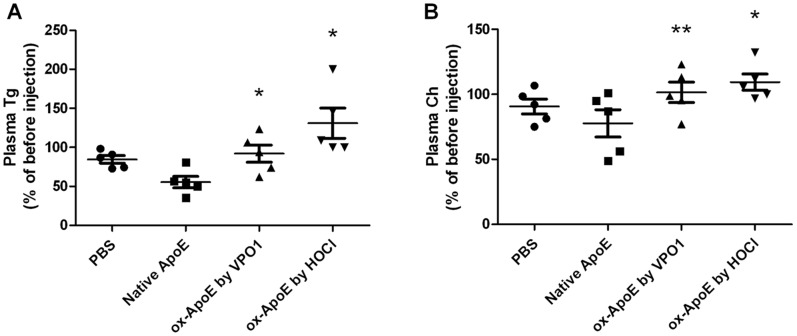
Results: Oxidized ApoE binds weaker to lipid emulsion particles, which mimic the large lipid complexes in vivo. In lipid efflux assay, oxidized ApoE showed reduced capability in efflux of lipids from foam cells. Mice administrated with oxidized ApoE via blood exhibited weaker clearance ability of plasma lipids.

Conclusions: Our data suggest that VPO1 is a new mediator regulating lipid homeostasis, implying a role in genesis and development of atherosclerosis.


Conflict of interest statement

Competing Interests: The authors have declared that no competing interests exist.

Figures

Figure 1
Figure 1. Localization of VPO1 and ApoE in atherosclerotic lesion.
Immunofluorescence staining was carried out for detection of ApoE (A) and VPO1 (B) using sections from mouse atherosclerotic lesions. C. Merged image of A and B. D. Nuclei were visualized with Hoechst staining (blue). E. Bright-field image. F. Merged image of A, B, D, and E. Scale bar, 20 µm. Magnification: x400.
Figure 2
Figure 2. Immunoblot analysis of ApoE oxidized by VPO1.
A. VLDL was oxidized by MPO or VPO1 as described in Materials and Methods. The oxidized lipoproteins were separated by SDS-PAGE and transferred to PVDF membrane. The blot was analyzed by using anti-ApoE antibody and visualized using chemiluminescence. B. The same as in A, but the samples were separated by using native PAGE. C. rApoE was oxidized by VPO1 or reagent HOCl. Blot was analyzed as in A.
Figure 3
Figure 3. Loss of free amino groups and tryptophan residues in oxidized ApoE.
A. Loss of free amino groups in oxidized ApoE. ApoE was oxidized by VPO1/H2O2/Cl, MPO/H2O2/Cl, or HOCl for 3 hrs at 37°C (n = 3). Unmodified amino groups were quantified with the method using trinitrobenzene sulfonic acid as described in Materials and Methods. Absorbance was measured at 340 nm. B. Loss of tryptophan residues following ApoE oxidation. ApoE was incubated with VPO1/H2O2/Cl, MPO/H2O2/Cl or HOCl for 3 hrs at 37°C (n = 3) as in A. Fluorescence from free tryptophan residue (emission 335 nm/excitation 280 nm) was quantified with a BioTek Microplate Reader. Trp: tryptophan. Data are shown as means ± SEM; *p<0.05 vs. native ApoE; n = 3.
Figure 4
Figure 4. Ox-ApoE binds weakly to emulsion particles.
A. Ox-ApoE binding with triglyceride-PC emulsion particles. Native and ox-ApoE were incubated with triglyceride-PC emulsion particles at room temperature for 1 hr with gentle shaking. Emulsion particles and lipid-free buffer were segregated by centrifugation. Equal amount of samples from top and lower layers (contain lipid-bound ApoE and free ApoE, respectively) were carried out for Western blot analysis using anti-ApoE antibody. B. Ox-ApoE binding with PC emulsion particles. Native and ox-ApoE were incubated with PC emulsion particles at room temperature for 1 hr with gentle shaking. Emulsion particles and lipid-free buffer were segregated by centrifugation as in A. Equal amount of samples from top and lower layers (contain lipid-bound ApoE and free ApoE, respectively) were carried out for Western blot analysis using anti-ApoE antibody. C. Quantification of ApoE and ox-ApoE binding to triglycerides-PC emulsion particles from data in A. The ratio was calculated as emulsion particle-bound ApoE/free ApoE. D. Quantification of ApoE and ox-ApoE binding to PC emulsion particles from data in B. The ratio was calculated as emulsion particle-bound ApoE/free ApoE. Data are shown as means ± SEM; *p<0.05. n = 3.
Figure 5
Figure 5. Effect of ox-ApoE on triglyceride, cholesterol and PC efflux from foam cells.
A. Cellular triglyceride efflux by native and ox-ApoE. B. Cellular cholesterol efflux by native and ox-ApoE. C. Cellular PC efflux by native and ox-ApoE. D. Detection of triglycerides in supernatant. E. Detection of cholesterol in supernatant. F. Detection of PC in supernatant. THP-1 cells were loaded with lipids as described in Materials and Methods. Efflux of triglycerides, cholesterol, and PC was initiated by addition of native or ox-ApoE in cultured foam cells. After treatment of 9 hrs, triglycerides, cholesterol, and PC in supernatant were measured by the respect kit. After treatment of 24 hrs, the cellular triglycerides, cholesterol, and PC were measured with the respect kit. Tg: triglycerides; Ch: cholesterol; PC: phosphatidylcholine. Data are shown as means ± SEM; *p<0.05 vs. PBS group. n = 3.
Figure 6
Figure 6. Effect of ox-ApoE on the transportation of plasma triglycerides and cholesterol.
A. Changes of plasma triglycerides by administration of native and ox-ApoE. B. Changes of plasma cholesterol by administration of native and ox-ApoE. Plasma triglyceride and cholesterol concentrations were measured 1 hr before and after of ApoE administration. *p<0.05 vs. native ApoE. **p<0.05 vs. native ApoE (paired t-test). n = 5.

References

    1. Choy PC, Siow YL, Mymin D, Karmin O (2004) Lipids and atherosclerosis. Biochem Cell Biol 82: 212–224. - PubMed
    1. Cox RA, Garcia-Palmieri MR (1990) Cholesterol, Triglycerides, and Associated Lipoproteins. In: Walker HK, Hall WD, Hurst JW, editors. Clinical Methods: The History, Physical, and Laboratory Examinations. 3rd ed. Boston. - PubMed
    1. Mahley RW, Innerarity TL, Rall SC Jr, Weisgraber KH (1984) Plasma lipoproteins: apolipoprotein structure and function. J Lipid Res 25: 1277–1294. - PubMed
    1. Mahley RW (1988) Apolipoprotein E: cholesterol transport protein with expanding role in cell biology. Science 240: 622–630. - PubMed
    1. Sing CF, Davignon J (1985) Role of the apolipoprotein E polymorphism in determining normal plasma lipid and lipoprotein variation. Am J Hum Genet 37: 268–285. - PMC - PubMed

Publication types