Review
. 2013 Mar;137(3):314-25.
doi: 10.5858/arpa.2012-0075-RA.

Making the diagnosis of frontotemporal lobar degeneration

Affiliations
Review

Making the diagnosis of frontotemporal lobar degeneration

Eileen H Bigio. Arch Pathol Lab Med. 2013 Mar.

Abstract

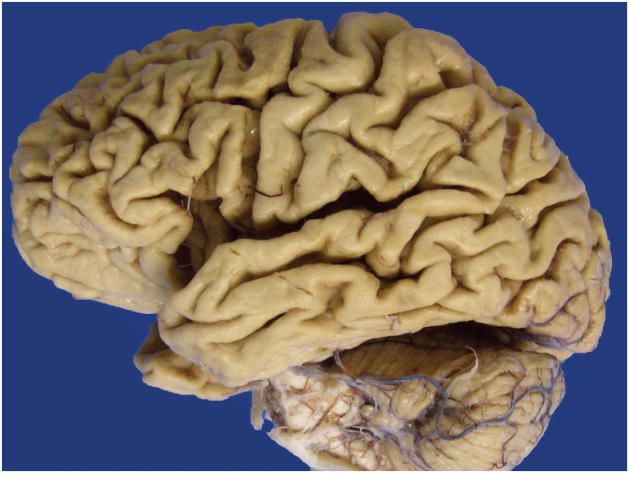
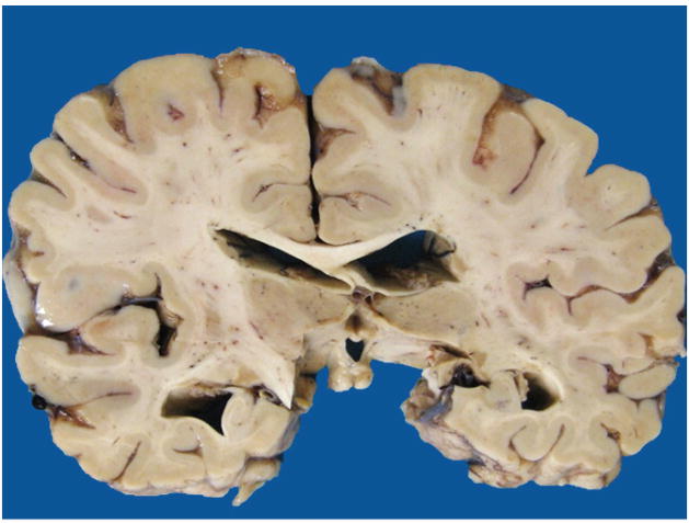
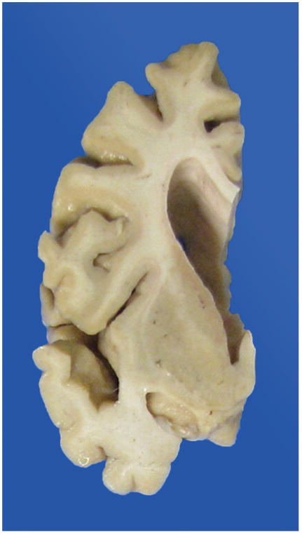
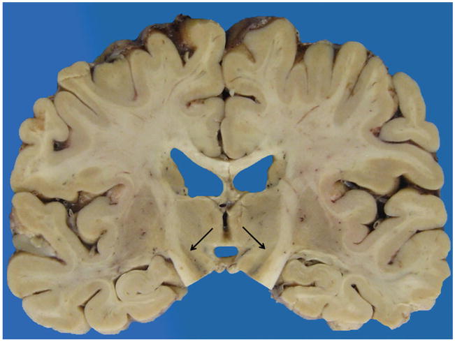
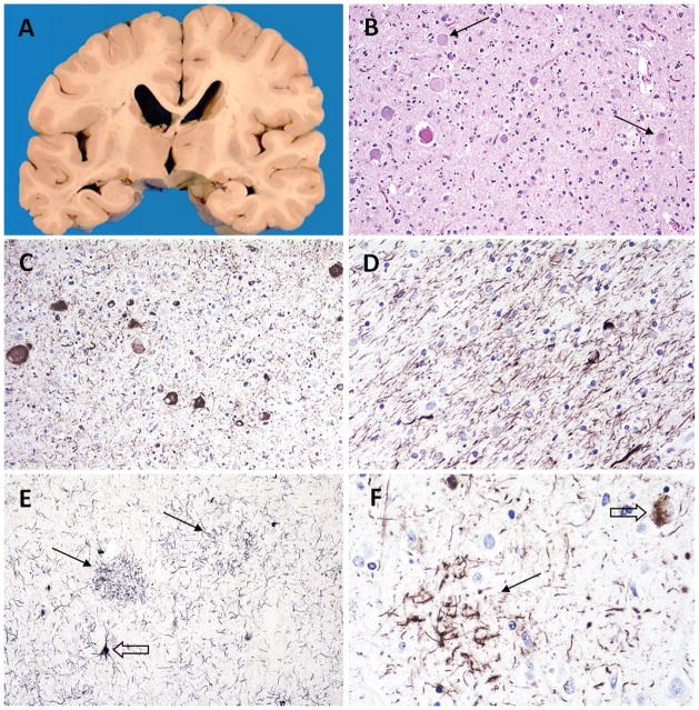
Context: Autopsy evaluation of the brain of a patient with frontotemporal dementia (FTD) can be daunting to the general pathologist. At some point in their training, most pathologists learn about Pick disease, and can recognize Pick bodies, the morphologic hallmark of Pick disease. Pick disease is a type of frontotemporal lobar degeneration (FTLD), the general category of pathologic process underlying most cases of FTD. The 2 major categories of pathologic FTLD are tauopathies (FTLD-tau) and ubiquitinopathies (FTLD-U). Pick disease is one of the FTLD-tau subtypes and is termed FTLD-tau (PiD).

Objective: To "demystify" FTLDs, and to demonstrate that subtypes of FTLD-tau and FTLD-U can be easily determined by following a logical, stepwise, histochemical, and immunohistochemical investigation of the FTD autopsy brain.

Data sources: Previously published peer-reviewed articles.

Conclusions: The hope is that this article will be a useful reference for the general pathologist faced with performing a brain autopsy on a decedent with frontotemporal dementia.


Conflict of interest statement

The author has no relevant financial interest in the products or companies described in this article.

Figures

Figure 1
Figure 1
Generalized cortical atrophy.
Figure 2
Figure 2
Hippocampal atrophy, moderate.
Figure 3
Figure 3
Frontal and superior temporal gyrus atrophy greater than parietal and occipital atrophy.
Figure 4
Figure 4
Caudate atrophy, severe.
Figure 5
Figure 5
Subthalamic nucleus atrophy, severe.
Figure 6
Figure 6
Cerebellar dentate nucleus “indistinct.”
Figure 7
Figure 7
Nigral greater than locus pallor.
Figure 8
Figure 8
Illustration showing where to take 13 histologic sections.
Figure 9
Figure 9
Flowchart showing sequence of stains and immunostains used to make the diagnosis of a specific frontotemporal lobar degeneration. Abbreviations: Aβ, amyloid-β; AD, Alzheimer disease; aFTLD-U, atypical frontotemporal lobar degeneration; AGD, argyrophilic grain disease; ALS, amyotrophic lateral sclerosis; Amyg, amygdala; α-syn, α-synuclein; BG, basal ganglia; BIBD, basophilic inclusion body disease; Biel, Bielschowsky; CBD, corticobasal degeneration; Cbm, cerebellum; DN, dentate nucleus; FTD-3, frontotemporal dementia associated with chromosome 3; FTLD-FUS, frontotemporal lobar degeneration with fused in sarcoma proteinopathy; FTLD-ni, frontotemporal lobar degeneration with no inclusions; FTLD-tau, frontotemporal lobar degeneration with tauopathy; FTLD-TDP, frontotemporal lobar degeneration with TDP-43 proteinopathy; FTLD-UPS, frontotemporal lobar degeneration with involvement of the ubiquitin proteasome system; FUS, fused in sarcoma protein; Hippo, hippocampus; IHC, immunohistochemistry; IP, inferior parietal region; LBD, Lewy body disease; Med, medulla; MFG, middle frontal gyrus; Mid, midbrain; MSTD, multiple system tauopathy with dementia; NIFID, neuronal intermediate filament inclusion dementia; PiD, Pick disease; PSP, progressive supranuclear palsy; SC, spinal cord; STG, superior temporal gyrus; Str, striatum; TDP-43, transactive response deoxyribose nucleic acid binding protein of 43 kDa; Thal, thalamus; Thio, thioflavin-S; TPSD, tangle predominant senile dementia; Ub, ubiquitin; WMT-GGI, white matter tauopathy with globular glial inclusions.
Figure 10
Figure 10
Case 1: FTLD-tau (PiD) pathology. A, Coronal section at the level of the posterior hippocampus, showing hippocampal atrophy that is severe on the left and mild on the right. B, Pick bodies in dentate gyrus. C, Some dentate gyrus Pick bodies are immunopositive with ubiquitin. D, All Pick bodies are labeled with tau. E, Severe cortical neuronal loss and gliosis; ballooned neurons (inset). F, Cortical Pick bodies (arrows) and glial inclusions labeled with tau (open arrows) (hematoxylin-eosin, original magnifications ×400 [B], ×50 [E], and ×200 [E, inset]; Dako polyclonal ubiquitin antibody [Carpinteria, California], original magnification ×600 [C]; AT8 antibody [Pierce Endogen, Rockford, Illinois], original magnifications ×600 [D] and ×400 [F]).
Figure 11
Figure 11
Case 2: FTLD-tau (CBD) pathology. A, Coronal section at the level of the posterior hippocampus, showing no cortical asymmetry and moderate ventricular dilatation. B, Ballooned neurons in motor cortex; arrows point to two. C, Ballooned neurons and cortical threads labeled with tau. D, White matter threads and glial inclusions labeled with tau. E, Astrocytic plaques (arrows), neuronal inclusions (open arrow), and cortical threads stain positively with silver stain. F, Astrocytic plaque (arrow) and neuronal inclusion (open arrow) labeled with tau (hematoxylin-eosin, original magnification ×200 [B]; AT8 [Pierce Endogen, Rockford, Illinois], original magnifications ×200 [C] and ×400 [D and F]; Gallyas stain, original magnification ×200 [E]).
Figure 12
Figure 12
Case 3: FTLD-tau (PSP) pathology. A, Coronal section at the level of the anterior hippocampus, showing mild to moderate cortical atrophy, severe ventricular dilatation, severe atrophy of the head of the caudate (white arrows), and mild atrophy and yellowish discoloration of the globus pallidus (black arrows). B, Globus pallidus with tau-positive neuronal inclusion (arrow) and several tau-positive tufted astrocytes (open arrow). C, Cerebellar dentate nucleus showing moderate neuronal loss and gliosis; synaptophysin highlights grumose degeneration (arrows) in dentate nucleus (inset). D, Rare tau-positive neurons in dentate nucleus. E, Tau-positive neurons and tufted astrocytes in cortex; silver-positive tufted astrocyte (inset). F, Tau-positive tufted astrocytes in putamen. G, Tau-positive neurons in subthalamic nucleus (AT8 [Pierce Endogen, Rockford, Illinois], original magnifications ×200 [B and G], ×100 [D and F], and ×50 [E]; hematoxylin-eosin, original magnification ×200 [C]; Dako [Carpinteria, California] monoclonal synaptophysin antibody SY38, original magnification ×200 [C, inset]; Gallyas stain, original magnification ×200 [E, inset]).
Figure 13
Figure 13
Case 4: FTLD-TDP pathology. Marked asymmetry of cortical atrophy, with severe right (A) and mild to moderate left (B) frontal, temporal, and parietal atrophy, demonstrated in coronal section at the level of the anterior hippocampus (C), which also shows severe right and moderate left ventricular dilatation. Note relative sparing of motor and sensory gyri. TDP-43–positive cytoplasmic and intranuclear inclusions and short dystrophic neurites consistent with harmonized FTLD-TDP type A (D). A different case, showing FTLD-TDP type B pathology in cortex, with predominantly granular cytoplasmic inclusions (E). FTLD-TDP type B pathology in dentate gyrus. Note that antibody to normal TDP-43 highlights normal nuclear TDP-43 in neurons that do not have inclusions (F). Last, harmonized FTLD-TDP type C pathology (G) (phosphorylated TDP-43 antibody S409/410-2 [Cosmo Bio, Carlsbad, California], original magnifications ×600 [D] and ×200 [G]; antibody to normal TDP-43 [ProteinTech, Chicago Illinois], original magnifications ×400 [E and F]).
Figure 14
Figure 14
Case 5: FTLD-FUS (BIBD) pathology. Asymmetry of frontal cortex atrophy with moderate to severe right (A) and mild left (B) frontal atrophy. Basophilic inclusion in motor cortex neuron (C). Basophilic inclusions are unstained by ubiquitin (D) but are strongly labeled with FUS antibody (E). The FUS antibody also highlights numerous cytoplasmic inclusions in superficial frontal and motor cortex that are not apparent on hematoxylin-eosin stains (F) (hematoxylin-eosin, original magnification ×400 [C]; Dako ubiquitin antibody [Dako, Carpinteria California], original magnification ×600 [D]; FUS antibody [ProteinTech, Chicago Illinois original magnifications ×600 [E] and ×400 [F]).
Figure 15
Figure 15
Graphic display of frontotemporal lobar degeneration (FTLD) pathologic subtypes, with currently known genetic associations. Abbreviations: aFTLD-U, atypical frontotemporal lobar degeneration; AGD, argyrophilic grain disease; ALS, amyotrophic lateral sclerosis; BIBD, basophilic inclusion body disease; CBD, corticobasal degeneration; CTE, chronic traumatic encephalopathy; DLDH, dementia lacking distinctive histology; FTD-3, frontotemporal dementia associated with chromosome 3; FTLD-FUS, frontotemporal lobar degeneration with fused in sarcoma proteinopathy; FTLD-ni, frontotemporal lobar degeneration with no inclusions; FTLD-tau, frontotemporal lobar degeneration with tauopathy; FTLD-TDP, frontotemporal lobar degeneration with TDP-43 proteinopathy; FTLD-UPS, frontotemporal lobar degeneration with involvement of the ubiquitin proteasome system; FUS, fused in sarcoma protein; MSTD, multiple system tauopathy with dementia; NIFID, neuronal intermediate filament inclusion dementia; PiD, Pick disease; PSP, progressive supranuclear palsy; TDP-43, transactive response deoxyribose nucleic acid binding protein of 43 kDa; TPSD, tangle predominant senile dementia; Ub, ubiquitin; WMT-GGI, white matter tauopathy with globular glial inclusions; 3R, 3 microtubule-binding repeats, 4R, 4 microtubule-binding repeats.

References

    1. Mirra SS, Hart MN, Terry RD. Making the diagnosis of Alzheimer’s disease: a primer for practicing pathologists. Arch Pathol Lab Med. 1993;117(2):132–144. - PubMed
    1. Mirra SS, Heyman A, McKeel D, et al. The Consortium to Establish a Registry for Alzheimer’s Disease (CERAD), II: standardization of neuropathological assessment of Alzheimer’s disease. Neurology. 1991;41(4):479–486. - PubMed
    1. Braak H, Braak E. Neuropathological stageing of Alzheimer-related changes. Acta Neuropathol. 1991;82(4):239–259. - PubMed
    1. Hyman BT, Trojanowski JQ. Consensus recommendations for the postmortem diagnosis of Alzheimer disease from the National Institute on Aging and the Reagan Institute Working Group on diagnostic criteria for the neuropathological assessment of Alzheimer disease. J Neuropathol Exp Neurol. 1997;56(10):1095–1097. - PubMed
    1. Montine TJ, Phelps CH, Beach TG, et al. National Institute on Aging-Alzheimer’s Association guidelines for the neuropathologic assessment of Alzheimer’s disease: a practical approach. Acta Neuropathol. 2012;123(1):1–11. - PMC - PubMed

Publication types

MeSH terms

LinkOut - more resources