Review
. 2013:82:745-74.
doi: 10.1146/annurev-biochem-072611-130030. Epub 2013 Feb 28.

Systemic amyloidoses

Affiliations
Review

Systemic amyloidoses

Luis M Blancas-Mejía et al. Annu Rev Biochem. 2013.

Abstract

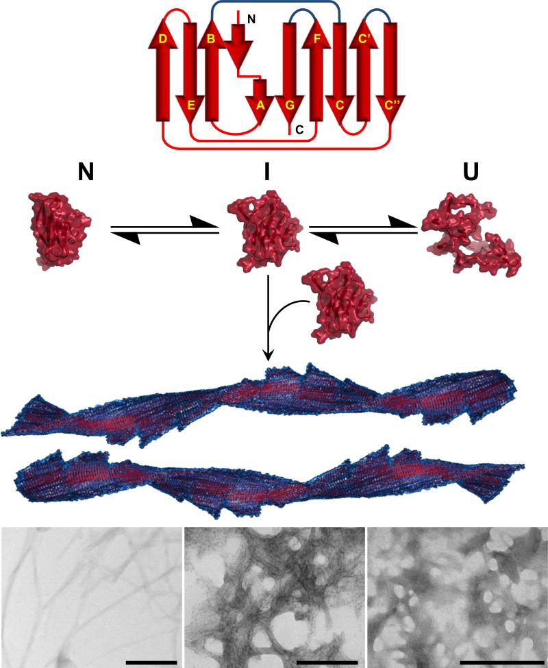
The amyloidoses are a group of protein misfolding diseases in which the precursor protein undergoes a conformational change that triggers the formation of amyloid fibrils in different tissues and organs, causing cell death and organ failure. Amyloidoses can be either localized or systemic. In localized amyloidosis, amyloid deposits form at the site of precursor protein synthesis, whereas in systemic amyloidosis, amyloid deposition occurs distant from the site of precursor protein secretion. We review the type of proteins and cells involved and what is known about the complex pathophysiology of these diseases. We focus on light chain amyloidosis to illustrate how biochemical and biophysical studies have led to a deeper understanding of the pathogenesis of this devastating disease. We also review current cellular, tissue, and animal models and discuss the challenges and opportunities for future studies of the systemic amyloidoses.


Figures

Figure 1
Figure 1
Amyloid fibril structure showing conversion from natively folded protein to partially unfolded intermediate to amyloid fibril. Electron microscopy images show different morphologies of amyloid fibrils.
Figure 2
Figure 2
Light chain (AL) amyloidosis pathology A) clonal expansion of plasma cells secreting light chain dimers that deposit in vital organs as amyloid fibrils B) Immunoglobulin structure showing two identical heavy chains (HC) and two identical light chains (LC) linked together via covalent interchain disulfide bonds.
Figure 3
Figure 3
(a) X-ray structure of κIgG antibody (PDB code: 1IGT). Two heavy chains (green), and two light chains (gold) assemble in a heterotetramer. (b) Structure of full length light chain (PDB code: 1IGT, chain A). Both the CL (red) and VL (yellow) domains present a characteristic immunoglobulin fold. (c) Structural alignment of X-ray crystal structures of λVL 6aJL2 germline (gold) and κVL κ1 O18/O8JK2 (red) germline proteins (PDB code: 2W0K and 2Q20, respectively). The FR region shows a very similar structure while the CDRs (blue) show significant differences as a result of high variability in the sequence. κ and λ germline proteins present a highly conserved single tryptophan residue at position 35 (black sticks), in close proximity of the characteristic immunoglobulin disulfide bridge (gray sticks).
Figure 4
Figure 4
This morph animation show the transition between three unique dimer orientation present in the crystal structures of κI O18/O8 and AL-09, as well as NMR structure of κI Y87H in order to see and understand the structural differences between them. This animation does not represent genuine conformational changes. Surface representation of the residues in the monomer A involved in the κI dimer interface is colored in magenta while residues outside the dimer interface are shown in cyan. Monomer B (ribbons) of AL-09 (blue, PDB code: 2Q1E), κVL κI O18/O8 (gold, PDB code: 2Q20), κI Y87H (red, PDB code: 2KQM) show a ~90° and ~ 180° rotation to the monomer A from the canonical dimer interface respectively. In yellow ribbon is shown the differences in dimer structures as the hands on a clock moving in intervals of 90°. Interface residues Q3, Y36, F98 and Q100 (red sticks) show the biggest conformational differences between dimer interfaces.
Figure 5
Figure 5
Structural comparison between the N-Terminal region of (a) λVL proteins and (b) κVL proteins (PDB codes: 2W0K, 2W0L, 3B5G, 1CD0, 1PW3, 1PEW, 2CD0, 2Q20, 2KQN, 3CDY, 2Q1E, 3DVI, 2KQM, 3CDC, 3DVF respectively). Significant differences in the N-terminal region are observed. κVL proteins present more H-bond (cyan lines) interactions between β-strand A (gold) and the edges of β-strands B (green) and β-strand G (red). In both proteins β-strand A adopts a characteristic conformation known as “β-sheet switch”, which has been proposed to protect the two edge β-strands B and G from non-native intermolecular interactions that lead to aggregation. The topology of the N-terminal region in both germline sequences is similar and includes a β-bulge (residues 7-8) between two segments of β-strand A. Interestingly, the size of the β-bulge in κVL is greater than in λVL proteins, and κVL presents more contacts between β-strand A and the C-terminus and β-strand B than λVL. Red circles indicate the β-bulge motif in β-strand G. CDRs are at the bottom of the models.

References

    1. Sipe JD, Benson MD, Buxbaum JN, Ikeda S-I, Merlini G, et al. Amyloid fibril protein nomenclature: 2010 recommendations from the nomenclature committee of the International Society of Amyloidosis. Amyloid. 2010;17:101–04. - PubMed
    1. Chiti F, Dobson CM. Protein misfolding, functional amyloid, and human disease. Annu Rev Biochem. 2006;75:333–66. - PubMed
    1. Sunde M, Serpell LC, Bartlam M, Fraser PE, Pepys MB, Blake CC. Common core structure of amyloid fibrils by synchrotron X-ray diffraction. J Mol Biol. 1997;273:729–39. - PubMed
    1. Geddes AJ, Parker KD, Atkins EDT, Beighton E. “Cross-β” conformation in proteins. Journal of Molecular Biology. 1968;32:343–58. - PubMed
    1. Eanes ED, Glenner GG. X-ray diffraction studies on amyloid filaments. Journal of Histochemistry & Cytochemistry. 1968;16:673–77. - PubMed

Publication types