Review
. 2013:42:361-92.
doi: 10.1146/annurev-biophys-083012-130326. Epub 2013 Mar 1.

Influences of membrane mimetic environments on membrane protein structures

Affiliations
Review

Influences of membrane mimetic environments on membrane protein structures

Huan-Xiang Zhou et al. Annu Rev Biophys. 2013.

Abstract

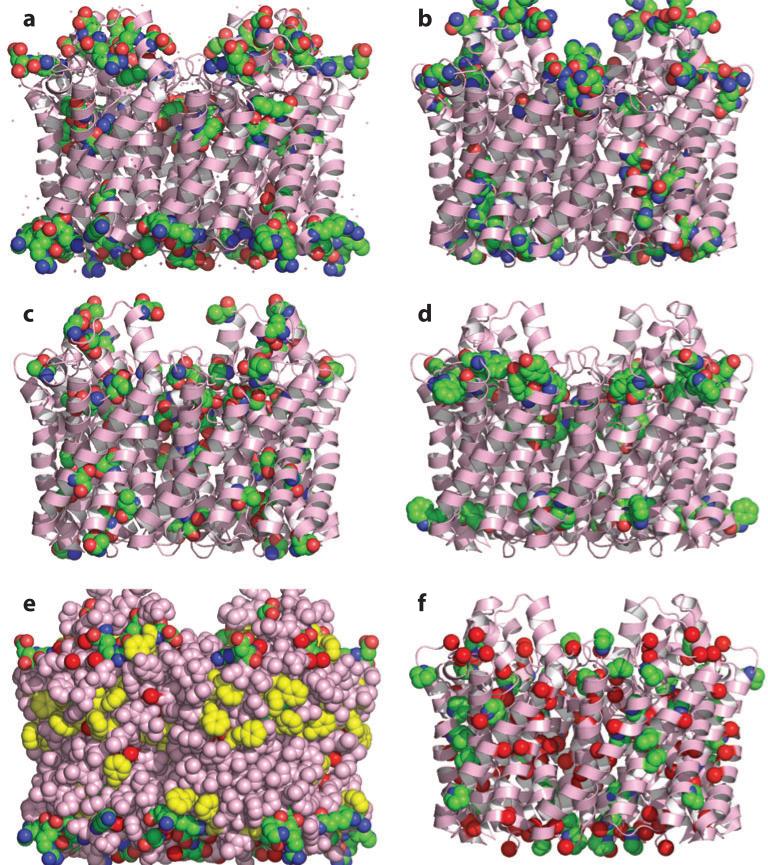
The number of membrane protein structures in the Protein Data Bank is becoming significant and growing. Here, the transmembrane domain structures of the helical membrane proteins are evaluated to assess the influences of the membrane mimetic environments. Toward this goal, many of the biophysical properties of membranes are discussed and contrasted with those of the membrane mimetics commonly used for structure determination. Although the mimetic environments can perturb the protein structures to an extent that potentially gives rise to misinterpretation of functional mechanisms, there are also many structures that have a native-like appearance. From this assessment, an initial set of guidelines is proposed for distinguishing native-like from nonnative-like membrane protein structures. With experimental techniques for validation and computational methods for refinement and quality assessment and enhancement, there are good prospects for achieving native-like structures for these very important proteins.


Figures

Figure 1
Figure 1
Comparison of M2 structures determined in different membrane mimetic environments. (a) Two detergent-based crystal structures of the transmembrane domain, 3BKD (pink) (91) and 3LBW (cyan; carrying the G34A mutation) (2), superimposed to 2L0J (green) (85), the solid-state NMR structure of the conductance domain in lipid bilayers. (b) Two micelle-based solution NMR structures of the conductance domain, 2RLF (yellow) (81) and 2KWX (blue; carrying the V27A mutation) (75), superimposed to 2L0J.
Figure 2
Figure 2
Amino acid distribution in the transmembrane domain of the glycerol facilitator (1FX8) (29) shown as pink ribbon except in panel e, where the surface is emphasized by showing an all-atom representation (pink except for highlighted residues in yellow). For the space-filling atoms: carbon, green spheres; nitrogen, blue spheres; and oxygen, red spheres, unless otherwise noted. (a) Charged residues Asp, Glu, Arg, and Lys. (b) Highly polar residues His, Asn, and Gln. (c) Polar residues Ser and Thr. (d ) Polar aromatic residues Trp and Tyr. (e) Charged residues (green carbons), Phe (yellow carbons), and Gly Cα (red carbons). (f ) Gly Cα (red carbons) and Pro ring atoms (green carbons).
Figure 3
Figure 3
Bilayer-like lattices in crystals. (a) Aquaporin (2W1P) (27) lattice structure with a parallel orientation of tetramers and minimal contacts between tetramers. (b) Antiparallel packing of M2 transmembrane domain tetramers (3BKD) (91), highlighting contacts between charged residues (space filling).
Figure 4
Figure 4
Nonbilayer lattices. (a) Leukotriene C4 synthase (2UUH) (60), showing a tetrahedral arrangement of trimers. (b) Estrone sulfatase (1P49) (38), showing helix pairs from three monomers interacting in a nearly orthogonal arrangement.
Figure 5
Figure 5
Electrostatic crystal contacts. (a) Crystal contacts in 2QTS (42), between the transmembrane domain of one ASIC1 trimer (green) and the water-soluble domain of a neighboring trimer (tan). (b) Enlarged view of the boxed region in panel a, highlighting the electrostatic contacts. (c) Crystal contacts in 2JLN (100), involving multiple charged residues at the interface between different Mhp1 molecules. (d) Intertrimer interactions between charged residues (Arg117, D142, and E144) in the apparently displaced last helices by neighboring FLAP (5-lipoxygenase-activating protein) trimers (2Q7M) (25).
Figure 6
Figure 6
Hydrophobic crystal contacts. (a) Two antiparallel tetramers of the M2 transmembrane domain in 3C9J (91), showing splayed helices. (b) Parallel (intratrimer; all green) and antiparallel (intertrimer; green versus yellow) arrangements of rhomboid protease monomers in the 2IC8 bilayer-like lattice (98). (c) Space-filling view of two antiparallel 2IC8 monomers at the intertrimer interface.
Figure 7
Figure 7
Hydrophobic dimensions of transmembrane domains. (a) Gloeobacter violaceus pentameric ligand-gated ion channel (GLIC) (3EAM) (5) displaying a substantial lipid annulus along with an appropriate hydrophobic thickness. (b) P2X4 receptor (4DW1) (35) with a thin hydrophobic dimension resulting from kinked and highly tilted helices. (c) Surface representation of 4DW1 highlighting the significant opening from the hydrophobic environment into the channel pore.
Figure 8
Figure 8
Weak hydrophobic environments in crystal lattices. (a) Rhomboid protease monomers (2IC8) (98) surrounded by detergent and water molecules (carbon, green spheres; oxygen, red spheres). (b) Leukotriene C4 synthase (2UUH) (60) displaying water molecules throughout the surface of the structure. (c) PepTSo (2XUT) (68), with cavities showing poor packing of the last two helices with the rest of the transmembrane domain.
Figure 9
Figure 9
Detergent molecules in and around transmembrane (TM) domains. (a) Proton-translocating pyrophosphatase (4A01) (53), with detergents appropriately lining the protein surface. (b) M2 TM domain (3BKD) (91), with detergents in the pore and between helices. Detergents wedged between helices in (c) the NCS2 transporter (3QE7) (56), (d) a fucose transporter (3O7P) (19), and (e) a sodium/calcium exchanger (3V5U) (52). (f) Putative sulfate permease (3TX3) (55), with detergents oriented randomly around TM helices.
Figure 10
Figure 10
Potential connection between helix splaying and weak lateral pressure. (a) Zinc transporter (3H90) (57), with two-helix bundles separated by a large gulf and cavities within the bundles. (b) Lipid flippase (3B60) (99), with a smaller opening between the helix bundles that may be required for protein function. In panels a and b, charged residues are displayed to delimit the hydrophobic thickness.
Figure 11
Figure 11
Different pore sizes and helix tilts in M2 structures. (a) 2RLF (81), conductance domain in DHPC (dihexanoyl phosphatidylcholine) micelles. (b) 2L0J (85), conductance domain in lipid bilayers. (c) 2KQT, transmembrane domain in lipid bilayers (6, 41). Two horizontal lines delimit the amantadine (AMT) binding site in the channel pore.
Figure 12
Figure 12
KdpD histidine kinase receptor (2KSF) (61) with two short helices. (a) Two exposed Gly residues (red spheres). (b) Amide protons (yellow spheres) displaying significant hydrogen/deuterium exchange. (c) Exposed hydrophilic side chains.
Figure 13
Figure 13
ArcB histidine kinase receptor (2KSD) (61). (a) Curved helix with amide hydrogen/deuterium exchange sites highlighted by yellow spheres. (b) Exposed hydrophilic side chains. (c) Exposed Gly residue (red sphere).
Figure 14
Figure 14
Stabilization of helix-helix packing by interhelical hydrogen bonds between side chains. (a) BNip3 dimer (2KA2) (92) stabilized by two His173-Ser172 hydrogen bonds and close packing afforded by a GxxxG motif. (b) CD3 ζζ dimer (2HAC) (8) stabilized by two Tyr12-Thr17 hydrogen bonds and close packing of the Asp2 side chains.
Figure 15
Figure 15
The diacylglycerol kinase (DAGK) trimer (2KDC) (95) with outward curved and poorly packed helices. (a) Charged residues, including those among the N-terminal 28 residues. (b) Large interhelical cavities. (c) Multiple Ala motifs not used for helix packing. In panels b and c, the N-terminal 28 residues are not displayed for clarity.
Figure 16
Figure 16
Different orientations of the amphipathic helices in two structures of the phospholamban pentamer. (a) 1ZLL (71). (b) 2KYV (96). For 2KYV, charged residues are shown in one amphipathic helix to indicate that it has an appropriate helical rotation.
Figure 17
Figure 17
Bacteriorhodopsin structures. (a) Electron crystallography (EC) structure of bacteriorhodopsin (2BRD) (33) in the native purple membrane environment, viewed down the bilayer normal to highlight lipids in the matrix surrounding the trimeric protein (one trimer and other monomers shown). (b) View into the bilayer plane, showing lipids surrounding a trimer. (c) Comparison of the EC structure (yellow) to an X-ray crystal structure (2NTU, orange) (50).
Figure 18
Figure 18
Trimeric structures from the MAPEG (membrane-associated proteins in eicosanoid and glutathione metabolism) superfamily, with charged residues highlighted in a monomer. (a) Electron crystallography structure of glutathione transferase 1 (2H8A) (40). (b) Comparison of 2H8A (green monomer) to the X-ray structure of FLAP (5-lipoxygenase-activating protein) (yellow and gray monomers, displaying charged residues) (2Q7M) (25).
Figure 19
Figure 19
Three structures determined by solid-state NMR (ssNMR) spectroscopy in lipid environments, with residues in the lipid interfacial region highlighted. (a) Gramicidin A (1MAG) (46). (b) MerF Hg transporter (2LJ2) (20). (c) Pf1 coat protein (2KSJ) (74).

References

    1. Abe K, Tani K, Nishizawa T, Fujiyoshi Y. Inter-subunit interaction of gastric H+,K+-ATPase prevents reverse reaction of the transport cycle. EMBO J. 2009;28:1637–43. - PMC - PubMed
    1. Acharya R, Carnevale V, Fiorin G, Levine BG, Polishchuk AL, et al. Structure and mechanism of proton transport through the transmembrane tetrameric M2 protein bundle of the influenza A virus. Proc. Natl. Acad. Sci. USA. 2010;107:15075–80. - PMC - PubMed
    1. Anfinsen CB. Principles that govern the folding of protein chains. Science. 1973;181:223–30. - PubMed
    1. Belrhali H, Nollert P, Royant A, Menzel C, Rosenbusch JP, et al. Protein, lipid water organization in bacteriorhodopsin: a molecular view of the purple membrane at 1.9Å resolution. Structure. 1999;7:909–17. - PubMed
    1. Bocquet N, Nury H, Baaden M, Le Poupon C, Changeux JP, et al. X-ray structure of a pentameric ligand-gated ion channel in an apparently open conformation. Nature. 2009;457:111–14. - PubMed

Publication types

LinkOut - more resources