Review
. 2013:42:289-314.
doi: 10.1146/annurev-biophys-050511-102349. Epub 2013 Feb 28.

Considering protonation as a posttranslational modification regulating protein structure and function

Affiliations
Review

Considering protonation as a posttranslational modification regulating protein structure and function

André Schönichen et al. Annu Rev Biophys. 2013.

Abstract

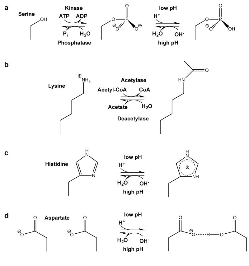
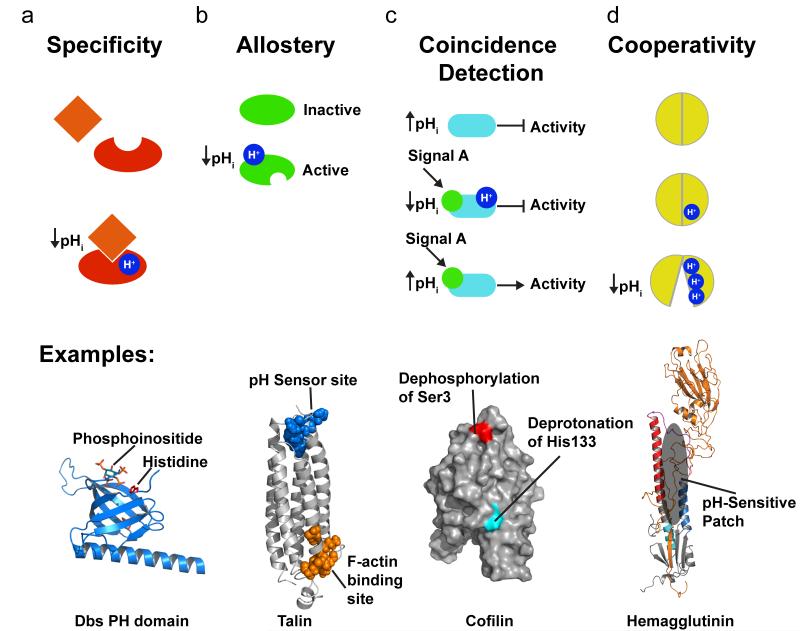
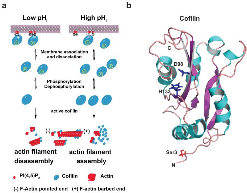
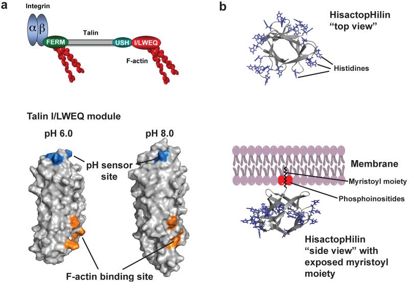
Posttranslational modification is an evolutionarily conserved mechanism for regulating protein activity, binding affinity, and stability. Compared with established posttranslational modifications such as phosphorylation or ubiquitination, posttranslational modification by protons within physiological pH ranges is a less recognized mechanism for regulating protein function. By changing the charge of amino acid side chains, posttranslational modification by protons can drive dynamic changes in protein conformation and function. Addition and removal of a proton is rapid and reversible and, in contrast to most other posttranslational modifications, does not require an enzyme. Signaling specificity is achieved by only a minority of sites in proteins titrating within the physiological pH range. Here, we examine the structural mechanisms and functional consequences of proton posttranslational modification of pH-sensing proteins regulating different cellular processes.


Figures

Figure 1
Figure 1
Examples of amino acid post-translational modifications associated with changes in charge. (a): Phosphorylation of e.g. serines (shown), threonines, tryptophans and histidines leads to addition of a negative charge at weakly basic conditions (151). (b): Lysine acetylation shields the lysine amino group e.g. to decrease the affinity to DNA (126), (c): Histidines can quickly abstract protons to shuttle protons or to function as a pH sensor site, (d): When pKa values are upshifted, protonation of carboxyl groups of glutamate or aspartate (shown) can lead to formation of new hydrogen bonds important for conformational changes of proteins.
Figure 2
Figure 2
Signaling modes regulated by pHi. (a) Protonation can regulate specificity as in protonation of a histidine in some but not all Rho GEFs that is required for stereospecific interaction with phosphoinositides. (b) An allosteric regulation mode occurs when protonation of a distinct site induces a conformational change a remote, as in talin that has an actin binding side ~40 Å away from the pH sensor (pdb code 2JSW). (c) In coincidence detection two distinctly and generally unrelated input signals as necessary for the output of protein function, as shown for cofilin that requires dephosphorylation of an N-terminal serine and deprotonation of a C-terminal histidine for increased activity. (d) Cooperativity occurs when several protonation sites act together with electrostatic coupling affecting titration and sometimes pKa shifts, as occurs with disrupted interactions of hemagglutinin HA1 domain with the HA2 domain.
Figure 3
Figure 3
Coincidence regulation of cofilin: (a) Model for coincidence detection of cofilin near the plasma membrane. At lower pHi < 7.2, the affinity of cofilin is higher, increasing binding to plasma membrane PI(4,5)P2 (red). At higher pHi > 7,2, less cofilin is bound to PI(4,5)P2, increasing the cytosolic pool of active cofilin if Ser3 is dephosphorylated. At higher pHi actin assembly is increased partly due to higher cofilin concentration. (b) Cartoon representation structure of human cofilin structure (pdb code: 1q8x) (109). The five α-helices are colored blue and the six β-sheets are colored purple. Side chains of Ser3, Asp98 and His133 are shown. N-terminal Ser3 is modified by phosphorylation. A salt bridge between Asp98 and His133 is formed under slight acidic conditions. His133 closely interacts with PI(4,5)P2 when doubly protonated (34).
Figure 4
Figure 4
Allosteric regulation of talin-actin binding by pH. (a) Domain organization of talin (upper panel), including an N-terminal FERM (domain that binds the β-subunit of integrin receptors and F-actin, a central rod domain (gray) and a C-terminal I/LWEQ (red) actin binding module that binding F-actin. F-actin binding by the I/LWEQ module but not by the FERM domain is pH sensitive with more binding at lower pH. Lower panel shows cartoon representation of C terminal actin-binding domain of talin. Residues E2337, E2342, H2418, D2482 form a pH sensor that induces pH-dependent conformations that allow actin binding only at lower pH (b) Cartoon representation of the histactopHilin structure and model of membrane attachment of hisactopHilin (pdb code: 1hcd). “Top view” shows that all histidines in loops in turns are equally distributed around the protein. However, viewed from the “side” these histidines point towards the cytoplasm, while the β-sheets point towards the membrane when the N-terminal myristoyl moiety is exposed partly by PI(4,5)P2.
Figure 5
Figure 5
Structural Regulation of hIAPP by pH. (a) IAPP sequences and segment propensity for fibril formation. The predicted energy for fibrillation of every six-residue segment of human IAPP, rat IAPP, and rat His18Arg IAPP are shown. Warmer colors represent a greater propensity for fibrillation, with red histogram bars represent hexapeptides that are predicted to form fibrils. Due to variations in the sequence, human IAPP has a much higher propensity to form fibrils than mouse IAPP. Mutation of Arg18 to His increases the propensity of rat IAPP to form fibrils. Graphs were generated using ZipperDB (http://services.mbi.ucla.edu/zipperdb/)(41) and modified from (153). (b) Potential model of pH dependent amyloid formation by IAPP. i) hIAPP, located in the insulin secretory granules, is bound to insulin b-chain (not shown) or to the membrane surface in an extended kinked helix conformation. ii) As IAPP is released from the acidic environment of the vesicle to the neurtral pH of the cytoplasma or extracellular space, His18 becomes deprotonated, weakening the insulin-hIAPP interaction and promoting insertion of hIAPP in the membrane by inducig a change in conformation. iii) Membrane associate hIAPP aggregates and, iv) undergoes further conformational changes leading to the formation of β-sheet-rich amyloid fibrils. Low pH structure: 2KB8 (103). Neutral pH structure: 2L86 (97). In both structures, the N terminus is located at the bottom of the figure. Figure adapted from (97; 150)
Figure 6
Figure 6
Low pH induces a large conformational change of the influenza HA2 protein (only one monomer is shown). (a) The structure of prefusion complex (left) of HA1 (gold, pdb code: 2hmg) and HA2 (multicolor, pdb code: 1htm) and the HA2 at the fusion pH (right) structure are shown as cartoon representations. In the prefusion complex the HA2 has a metastable conformation, and HA1 acts as a clamp to keep HA2 in this conformation. The hydrophobic fusion peptide (highlighted in orange) is buried in a hydrophobic core distant from the tip of the protein complex. Low pH leads to protonation of several charged residues throughout HA1 and HA2, and HA1 dissociates partly. This leads to a spontaneous conformational change of region B (magenta) that becomes an α-helix to form a new continuous helix A (blue), B and C (red). The fusion peptide moves ~100 Å from the core to the tip for insertion into the target membrane. Helix D (cyan) now packs against helix A, and a new hydrophobic core is formed. The C-terminus moves more towards the new N terminus and has more flexibility important for membrane fusion. (b) Schematic representation showing multimerization events of HIV gag and MA proteins as a function of pH (MA: matrix, CA: capsid, NC: nucleocapsid). Decreasing pH promotes myristoyl exposure membrane targeting and formation of multimers of both gag and MA protein, critical mediators for HI virus assembly. Structure figure reprinted with permisson from Jamil S. Saad (33).

References

    1. Alexov E, Mehler EL, Baker N, Baptista AM, Huang Y, et al. Progress in the prediction of pKa values in proteins. Proteins. 2011;79:3260–75. - PMC - PubMed
    1. Andreev OA, Engelman DM, Reshetnyak YK. pH-sensitive membrane peptides (pHLIPs) as a novel class of delivery agents. Mol Membr Biol. 2010;27:341–52. - PMC - PubMed
    1. Andrianantoandro E, Pollard TD. Mechanism of actin filament turnover by severing and nucleation at different concentrations of ADF/cofilin. Molecular cell. 2006;24:13–23. - PubMed
    1. Antonyuk SV, Trevitt CR, Strange RW, Jackson GS, Sangar D, et al. Crystal structure of human prion protein bound to a therapeutic antibody. Proc Natl Acad Sci U S A. 2009;106:2554–8. - PMC - PubMed
    1. Baddam S, Bowler BE. Conformationally gated electron transfer in iso-1-cytochrome c: engineering the rate of a conformational switch. J Am Chem Soc. 2005;127:9702–3. - PubMed

Publication types