

Histological predictors of outcome in ependymoma are dependent on anatomic site within the central nervous system
- PMID: 23452038
- PMCID: PMC8028973
- DOI: 10.1111/bpa.12050
Histological predictors of outcome in ependymoma are dependent on anatomic site within the central nervous system
Abstract
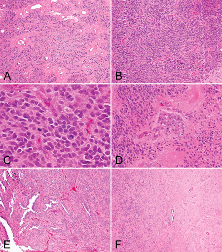
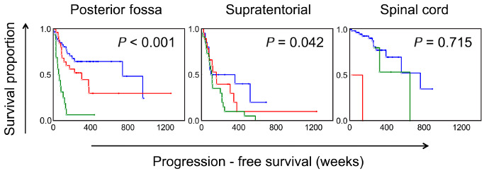
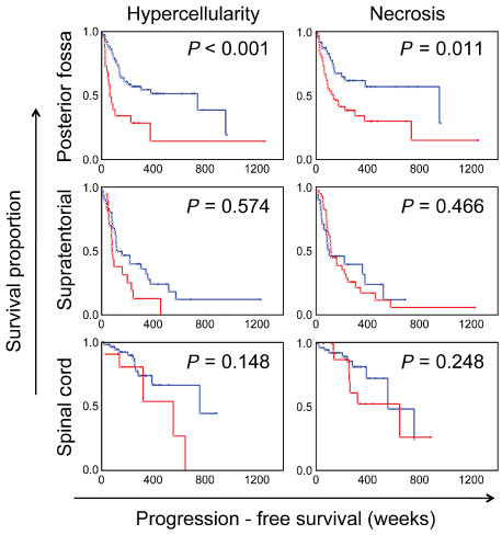
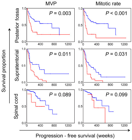
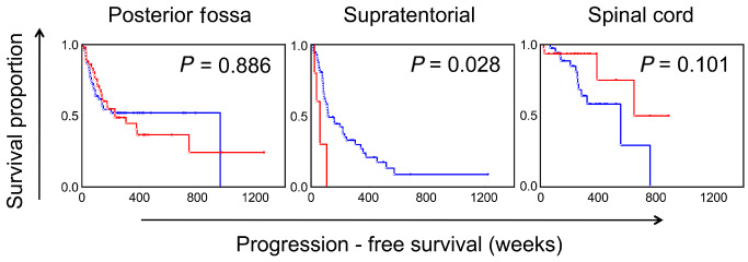
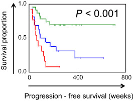
Ependymomas originate in posterior fossa (PF), supratentorial (ST) or spinal cord (SC) compartments. At present, grading schemes are applied independent of anatomic site. We performed detailed histological examination on 238 World Health Organization grade II and III ependymomas. Among PF ependymomas, the presence of hypercellular areas, necrosis, microvascular proliferation and elevated mitotic rate (all P < 0.01) were significantly associated with worse progression-free survival (PFS), while extensive ependymal canal formation was not (P = 0.89). Similar to the PF tumors, microvascular proliferation (P = 0.01) and elevated mitotic rate (P = 0.03) were significantly associated with worse PFS in the ST tumors. However, in contrast to PF tumors, extensive ependymal canals (P = 0.03) were associated with worse clinical outcome in ST ependymomas, but hypercellularity (P = 0.57) and necrosis (P = 0.47) were not. On multivariate Cox regression, after adjusting for relevant clinical variables, individual histological factors and a composite histological score remained significant among ST and PF ependymoma. In contrast to both PF and ST ependymoma, histological features were not found to be associated with PFS in SC tumors. Taken together, the clinical relevance of specific histological features in ependymoma appears to be related to the anatomic site of origin and suggests that site-specific grading criteria be considered in future classification systems.
Keywords: ependymoma; histology; progression-free survival.
© 2013 International Society of Neuropathology.
Figures
References
-
- Ernestus RI, Schroder R, Stutzer H, Klug N (1996) Prognostic relevance of localization and grading in intracranial ependymomas of childhood. Childs Nerv Syst 12:522–526. - PubMed
-
- Figarella‐Branger D, Civatte M, Bouvier‐Labit C, Gouvernet J, Gambarelli D, Gentet JC et al (2000) Prognostic factors in intracranial ependymomas in children. J Neurosurg 93:605–613. - PubMed
-
- Gilbert MR, Ruda R, Soffietti R (2010) Ependymomas in adults. Curr Neurol Neurosci Rep 10:240–247. - PubMed
Publication types
MeSH terms
Grants and funding
LinkOut - more resources
Full Text Sources
Other Literature Sources
