. 2013 May 15;452(1):57-66.
doi: 10.1042/BJ20121832.

Characterization of a pre-export enzyme-chaperone complex on the twin-arginine transport pathway

Affiliations

Characterization of a pre-export enzyme-chaperone complex on the twin-arginine transport pathway

Jennifer M Dow et al. Biochem J. .

Abstract

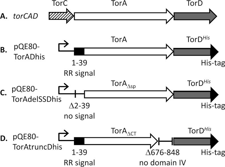
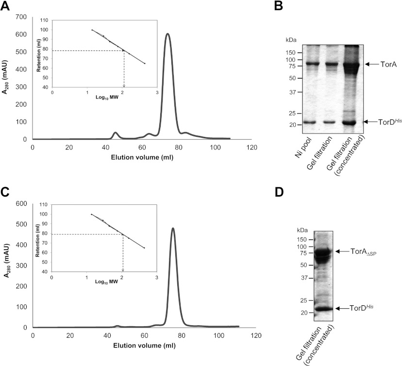
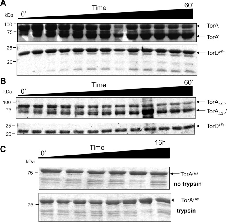
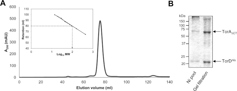
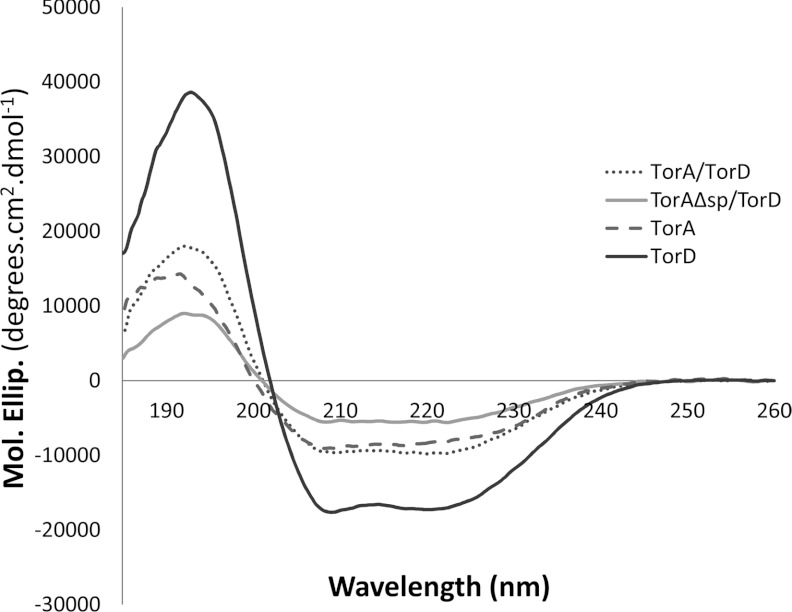
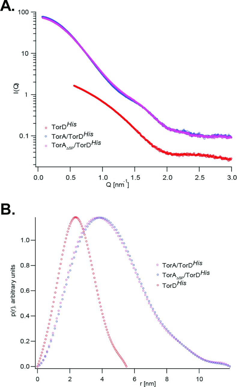
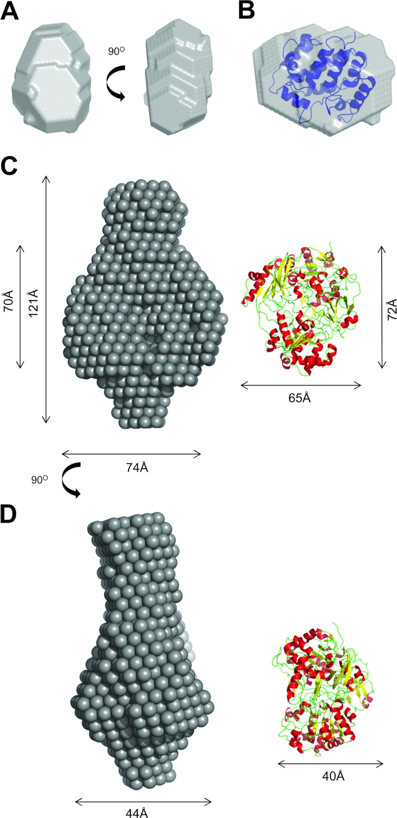
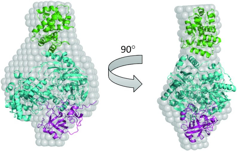
The Tat (twin-arginine translocation) system is a protein targeting pathway utilized by prokaryotes and chloroplasts. Tat substrates are produced with distinctive N-terminal signal peptides and are translocated as fully folded proteins. In Escherichia coli, Tat-dependent proteins often contain redox cofactors that must be loaded before translocation. Trimethylamine N-oxide reductase (TorA) is a model bacterial Tat substrate and is a molybdenum cofactor-dependent enzyme. Co-ordination of cofactor loading and translocation of TorA is directed by the TorD protein, which is a cytoplasmic chaperone known to interact physically with the TorA signal peptide. In the present study, a pre-export TorAD complex has been characterized using biochemical and biophysical techniques, including SAXS (small-angle X-ray scattering). A stable, cofactor-free TorAD complex was isolated, which revealed a 1:1 binding stoichiometry. Surprisingly, a TorAD complex with similar architecture can be isolated in the complete absence of the 39-residue TorA signal peptide. The present study demonstrates that two high-affinity binding sites for TorD are present on TorA, and that a single TorD protein binds both of those simultaneously. Further characterization suggested that the C-terminal 'Domain IV' of TorA remained solvent-exposed in the cofactor-free pre-export TorAD complex. It is possible that correct folding of Domain IV upon cofactor loading is the trigger for TorD release and subsequent export of TorA.


Figures

Figure 1
Figure 1. Tools for the isolation of TorAD complexes
(A) A cartoon representing the structure of the torCAD operon located at 22.8 min on the E. coli chromosome. The names of the protein products of the genes are given above the arrows. (B) An overexpression vector based on pQE80 (Qiagen) encoding full-length TorA and TorDHis. The natural transcriptional and translational coupling between torA and torD is maintained. (C) An expression vector encoding TorA lacking its entire Tat signal peptide comprising Asn2–Ala39 (TorAΔsp, TorA Δ2–39) and TorDHis. (D) An expression vector encoding TorA lacking its entire C-terminal Domain IV comprising Ile676–Ser848 (TorAΔCT, TorA Δ676–848) and TorDHis.
Figure 2
Figure 2. Isolation of a TorA–TorDHis and TorAΔSP–TorDHis complex
(A and C) TorDHis-containing fractions after metal chelate chromatography of cell extracts overproducing (A) TorA and TorDHis or (C) TorAΔSP and TorDHis were pooled, concentrated and applied to a HiLoad 16/60 Superdex 200 Prep Grade size-exclusion column. Eluted protein was monitored by measuring absorbance at 280 nm. The column was calibrated with the standard proteins ribonuclease (14 kDa), carbonic anhydrase (29 kDa), ovalbumin (44 kDa), conalbumin (75 kDa), aldolase (158 kDa) and ferritin (440 kDa), and the linear regression analysis is shown as inset boxes. R2=0.9976, y=−23.5x+126.83. MW, molecular mass. (B and D) SDS/PAGE analysis (12% gels) of (B) the concentrated fractions after metal chelate chromatography (Ni pool), and the non-concentrated and concentrated peak fractions from SEC of the TorA–TorDHis complex and (D) the concentrated peak fraction from SEC of the TorAΔSP–TorDHis complex. Molecular masses are indicated in kDa.
Figure 3
Figure 3. Limited trypsinolysis of the TorA–TorDHis and TorAΔSP–TorDHis complexes
The TorA–TorDHis (A) and TorAΔSP–TorDHis complexes (B) were isolated. Each purified complex was incubated with trypsin and 5 μl aliquots removed at 5 min intervals between 0 and 60 min, mixed with Laemmli buffer and immediately boiled to prevent any further digestion. (C) As a control, periplasmic mature TorAHis was purified and incubated with trypsin before 5 μl aliquots were removed at the following eight time points (0 min, 10 min, 20 min, 30 min, 1 h, 2 h, 4 h and 16 h). Concomitantly, mature TorAHis incubated without trypsin before aliquots were removed at the following six time points (0 min, 10 min, 30 min, 1 h, 4 h and 16 h). Samples were mixed with Laemmli buffer and immediately boiled. At the end of the time courses, the samples were analysed by SDS/PAGE (12% gels). Molecular masses are indicated in kDa.
Figure 4
Figure 4. Domain IV of TorA is not required for complex formation with TorD
(A) TorDHis-containing fractions after metal chelate chromatography of cell extracts overproducing TorAΔCT and TorDHis were pooled, concentrated and applied to a HiLoad 16/60 Superdex 200 Prep Grade size-exclusion column. Eluted protein was monitored by measuring absorbance at 280 nm. MW, molecular mass. (B) SDS/PAGE analysis (12% gels) of the pooled fractions after metal chelate chromatography (Ni pool), and the non-concentrated peak fraction following gel-filtration chromatography. Molecular masses are indicated in kDa.
Figure 5
Figure 5. CD spectra of purified TorAHis, TorDHis, and the TorA–TorDHis and TorAΔSP–TorDHis complexes
CD spectra (185–260 nm) were collected in quartz cells of 0.02 cm pathlength at 25°C with a scan speed of 10 nm/min, bandwidth of 1 nm, response of 2 s and data pitch of 0.2 nm. The buffer used was 50 mM Tris/H2SO4 (pH 7.5), 200 mM K2SO4 and 1 mM DTT, and protein concentrations were: TorA, 0.7 mg/ml; TorD, 1 mg/ml; TorA–TorDHis, 1 mg/ml; and TorAΔsp–TorDHis, 0.5 mg/ml.
Figure 6
Figure 6. SAXS characterization of TorDHis alone and TorDHis in complex with different forms of TorA
(A) SAXS results for TorDHis, and the TorA–TorDHis and TorAΔSP–TorDHis complexes. Q is the scattering vector and I represents intensity. (B) Distance distributions P(r) for TorDHis, TorA–TorDHis and TorAΔSP–TorDHis. All curves were normalized.
Figure 7
Figure 7. Ab initio modelling of TorDHis and the TorA–TorDHis complex
(A) Two different views of the ab intio model of TorD, generated using DAMMIN [37]. (B) The electron density for E. coli DmsD (PDB code 3CW0 [39]) docked into the SAXS-derived shape of TorDHis. (C) and (D) represent two different views of the ab intio model of the TorA–TorDHis complex. The X-ray structure of S. massilia TorA (PDB code 1TMO [32]) is shown to the right.
Figure 8
Figure 8. Rigid body modelling of the TorA–TorDHis complex
Rigid body modelling was conducted using the homologous component parts of the complex, i.e. TorD homologue DmsD (PDB code 3CW0, shown in green), and TorA (PDB code 1TMO) split into two parts: Domains I–III (i.e. amino acids 5–629; shown in cyan) and Domain IV (amino acids 631–798; shown in magenta). Modelling was conducted using SASREF [47]. The generated model is docked into the TorA–TorDHis ab intio model for comparison.

References

    1. Celedon J. M., Cline K. Intra-plastid protein trafficking: how plant cells adapted prokaryotic mechanisms to the eukaryotic condition. Biochim. Biophys. Acta. 2013;1833:341–351. - PMC - PubMed
    1. Palmer T., Berks B. C. The twin-arginine translocation (Tat) protein export pathway. Nat. Rev. Microbiol. 2012;10:483–496. - PubMed
    1. Berks B. C. A common export pathway for proteins binding complex redox cofactors? Mol. Microbiol. 1996;22:393–404. - PubMed
    1. Palmer T., Sargent F., Berks B. C. The Tat protein export pathway. In: Böck A., Curtiss R., Kaper J. B., Karp P. D., Neidhardt F. C., Nyström T., Slauch J. M., Squires C. L., Ussery D., editors. EcoSal–Escherichia coli and Salmonella: Cellular and Molecular Biology. Washington, DC: ASM Press; 2010. Chapter 4.3.2, http://www.ecosal.org.
    1. Sargent F. Constructing the wonders of the bacterial world: biosynthesis of complex enzymes. Microbiology. 2007;153:633–651. - PubMed

Publication types

MeSH terms

Substances

LinkOut - more resources