Review
. 2013 Mar;42(1):133-53.
doi: 10.1016/j.gtc.2012.11.008. Epub 2012 Dec 27.

Eosinophilic esophagitis

Affiliations
Review

Eosinophilic esophagitis

Evan S Dellon. Gastroenterol Clin North Am. 2013 Mar.

Abstract

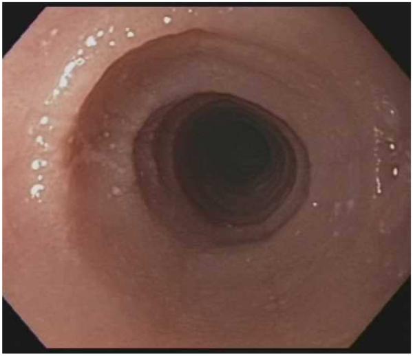
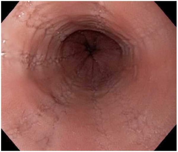
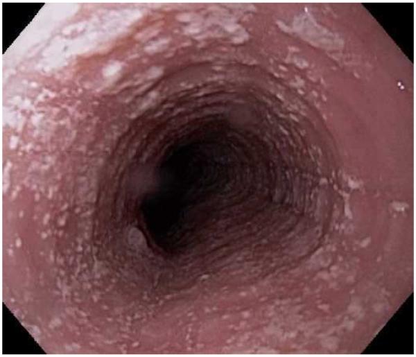
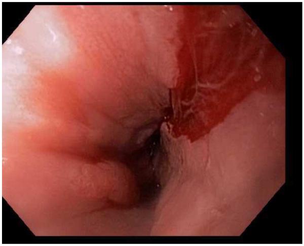
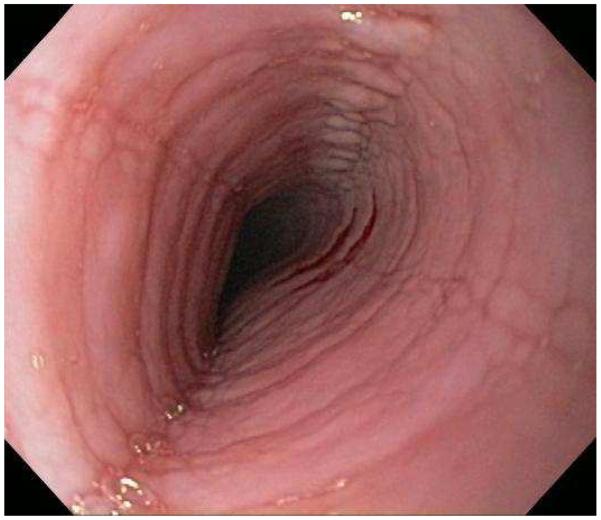
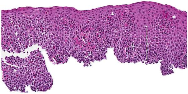
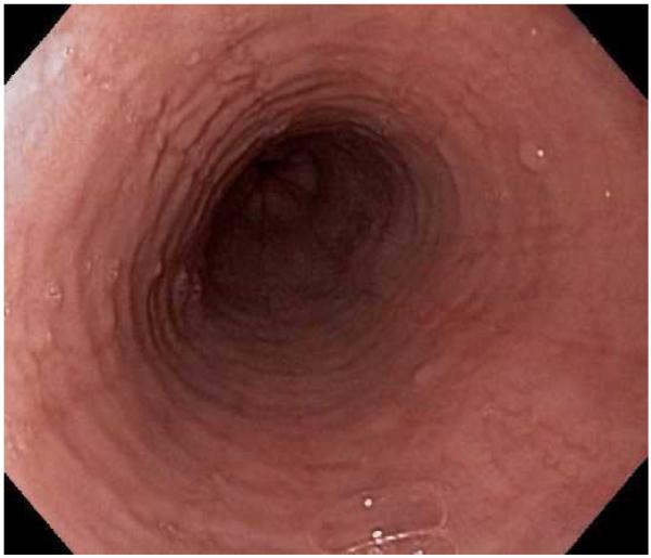
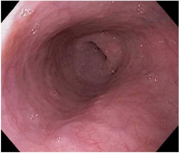
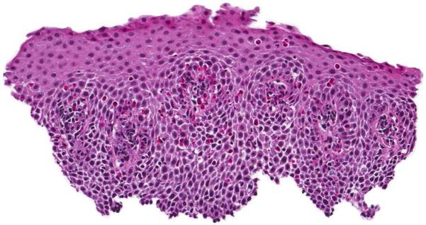
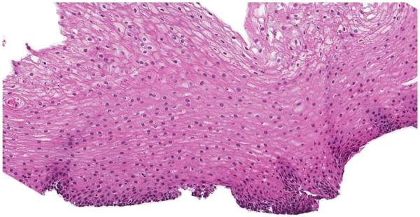
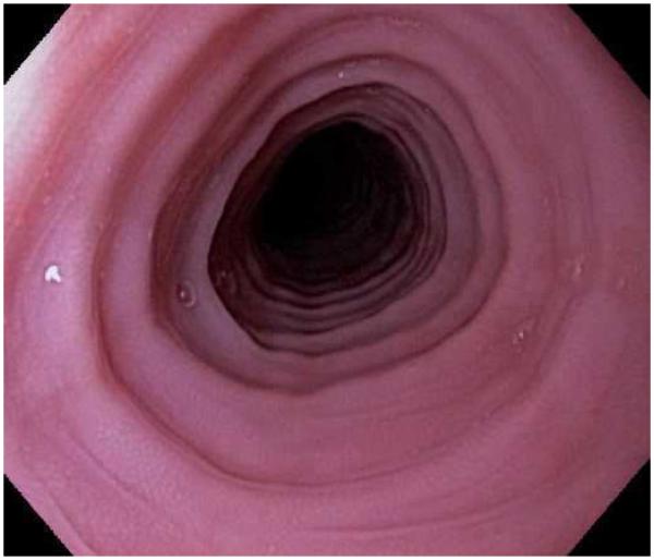
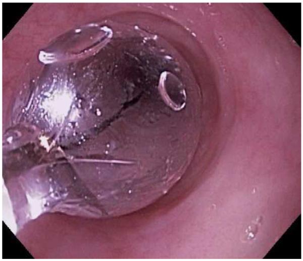
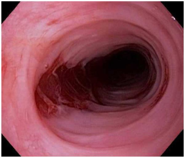
Eosinophilic esophagitis (EoE) is a chronic immune-mediated condition whereby infiltration of eosinophils into the esophageal mucosa leads to symptoms of esophageal dysfunction. EoE is encountered in a substantial proportion of patients undergoing diagnostic upper endoscopy. This review discusses the clinical, endoscopic, and histologic features of EoE and presents the most recent guidelines for its diagnosis. Selected diagnostic dilemmas are described, including distinguishing EoE from gastroesophageal reflux disease and addressing the newly recognized clinical entity of proton-pump inhibitor-responsive esophageal eosinophilia. Also highlighted is evidence to support both pharmacologic and nonpharmacologic treatments, including topical corticosteroids, dietary elimination therapy, and endoscopic dilation.


Figures

Figure 1
Figure 1
The range of endoscopic findings in EoE. (A) A patient with an acute food bolus impaction. (B) After the bolus is cleared, fixed esophageal rings are noted. (C) A patient with less prominent esophageal rings. (D) Felinization of the esophageal with rings that are transiently present. Congestion and loss of vascular marking area also noted. (E) A narrow caliber esophagus which prevented passage of an adult upper endoscope. Rings, congestion, and decreased vasculature are also present. (F) Linear furrows in the distal esophagus, associated subtle rings and decreased vasculature. (G) Prominent white plaques and exudates, as well as subtle rings and furrows. In this patient, brushings were negative for candida and biopsies showed multiple eosinophilic microabscesses. (H) Crêpe-paper mucosa where this tear was noted after uneventful passage of the endoscope. The mucosa is also congested with decreased vascularity. (I) A patient with multiple findings of rings, furrows, decreased vascularity, narrow caliber esophagus, and crêpe-paper mucosa.
Figure 1
Figure 1
The range of endoscopic findings in EoE. (A) A patient with an acute food bolus impaction. (B) After the bolus is cleared, fixed esophageal rings are noted. (C) A patient with less prominent esophageal rings. (D) Felinization of the esophageal with rings that are transiently present. Congestion and loss of vascular marking area also noted. (E) A narrow caliber esophagus which prevented passage of an adult upper endoscope. Rings, congestion, and decreased vasculature are also present. (F) Linear furrows in the distal esophagus, associated subtle rings and decreased vasculature. (G) Prominent white plaques and exudates, as well as subtle rings and furrows. In this patient, brushings were negative for candida and biopsies showed multiple eosinophilic microabscesses. (H) Crêpe-paper mucosa where this tear was noted after uneventful passage of the endoscope. The mucosa is also congested with decreased vascularity. (I) A patient with multiple findings of rings, furrows, decreased vascularity, narrow caliber esophagus, and crêpe-paper mucosa.
Figure 2
Figure 2
Esophageal biopsy specimen in EoE. This shows a brisk eosinophilic infiltration in the esophageal epithelium, as well as spongiosis (+), eosinophil degranulation (arrowhead), basal zone hyperplasia (bracket), and an eosinophilic microabscess (*).
Figure 3
Figure 3
(A) Endoscopic appearance of the esophagus prior to topical steroid therapy with rings, furrows, and degreased vascularity noted. (B) Appearance has completely normalized after topical steroid therapy. A similar improvement is noted on this patients biopsies before (C) and after (D) treatment.
Figure 3
Figure 3
(A) Endoscopic appearance of the esophagus prior to topical steroid therapy with rings, furrows, and degreased vascularity noted. (B) Appearance has completely normalized after topical steroid therapy. A similar improvement is noted on this patients biopsies before (C) and after (D) treatment.
Figure 4
Figure 4
Dilation for treatment of EoE. (A) Endoscopic appearance of a esophageal narrowing with a tight series of rings, as well as mucosal pallor, congestion, and decreased vasculature. (B) Through-the-scope balloon deployed and inflated to 10 mm. (C) Desired post-dilation effect with mucosal disruption.
Figure 4
Figure 4
Dilation for treatment of EoE. (A) Endoscopic appearance of a esophageal narrowing with a tight series of rings, as well as mucosal pallor, congestion, and decreased vasculature. (B) Through-the-scope balloon deployed and inflated to 10 mm. (C) Desired post-dilation effect with mucosal disruption.

References

    1. Liacouras CA, Furuta GT, Hirano I, et al. Eosinophilic esophagitis: Updated consensus recommendations for children and adults. J Allergy Clin Immunol. 2011;128:3–20.e6. - PubMed
    1. Landres RT, Kuster GG, Strum WB. Eosinophilic esophagitis in a patient with vigorous achalasia. Gastroenterology. 1978;74:1298–301. - PubMed
    1. Attwood SE, Smyrk TC, Demeester TR, Jones JB. Esophageal eosinophilia with dysphagia. A distinct clinicopathologic syndrome. Dig Dis Sci. 1993;38:109–16. - PubMed
    1. Straumann A, Spichtin HP, Bernoulli R, Loosli J, Vogtlin J. Idiopathic eosinophilic esophagitis: a frequently overlooked disease with typical clinical aspects and discrete endoscopic findings. Schweiz Med Wochenschr. 1994;124:1419–29. - PubMed
    1. Kelly KJ, Lazenby AJ, Rowe PC, Yardley JH, Perman JA, Sampson HA. Eosinophilic esophagitis attributed to gastroesophageal reflux: improvement with an amino acid-based formula. Gastroenterology. 1995;109:1503–12. - PubMed

Publication types

MeSH terms