. 2013 Mar 3:14:11.
doi: 10.1186/1471-2121-14-11.

Unlaid Xenopus eggs degrade by apoptosis in the genital tract

Affiliations

Unlaid Xenopus eggs degrade by apoptosis in the genital tract

Sho Iguchi et al. BMC Cell Biol. .

Abstract

Background: In several species with external fertilization, including frogs, laid unfertilized eggs were found to die by apoptosis outside of the animal body. However, there is no apparent reason for the externally laid eggs to degrade by this process, considering that apoptosis developed as a mechanism to reduce the damaging effect of individual cell death to the whole organism.

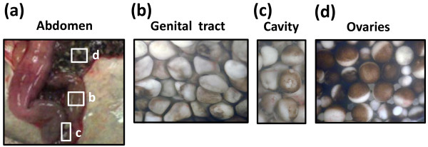
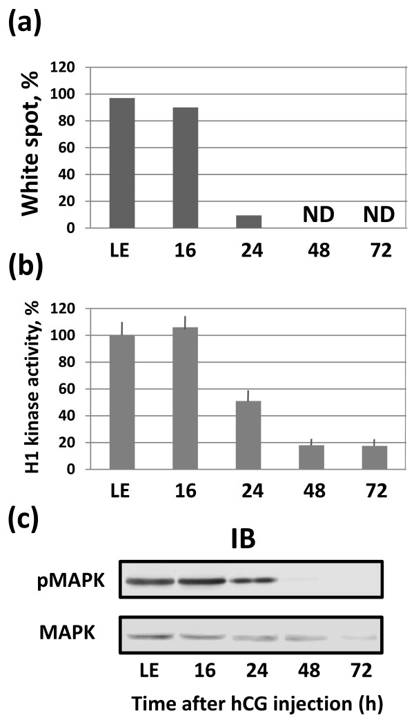
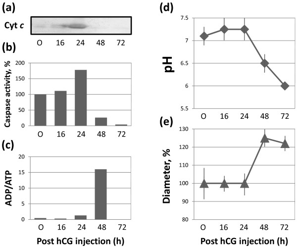
Results: Here, we demonstrate that a number of eggs are retained in the genital tract of the African clawed frog Xenopus laevis after gonadotropin-induced ovulation. The majority of these eggs exit meiotic arrest within 24 hours of hormone administration. Subsequently, post-meiotic eggs die in the frog genital tract by a well-defined apoptotic process. The hallmarks of egg degradation include prominent morphological changes, cytochrome c release, caspase activation, increase in ADP/ATP ratio, progressive intracellular acidification, egg swelling and all-out proteolysis of egg proteins. The sustained presence of post-apoptotic eggs in the genital tract of ageing frogs evidenced age-associated worsening of apoptotic clearance.

Conclusions: The direct observation of egg degradation in the Xenopus genital tract provides a clue to the physiological relevance of frog egg apoptosis. It works to eliminate the mature unlaid eggs retained in the animal body after ovulation. Our findings establish egg apoptosis as a major physiological process accompanying ovulation in frogs.


Figures

Figure 1
Figure 1
Ovulated eggs are retained in the frog body at different locations. The frog was dissected in 48 hours after hormonal stimulation (a) and the indicated fields of the abdomen (b, c, d) were observed microscopically.
Figure 2
Figure 2
Dynamics of egg degradation in the genital tract. Changes in the morphology of eggs retained in the frog genital tract over five days are shown in (a), the total volume of unlaid eggs in the uterus is presented in (b) and CBB staining of total egg proteins separated by SDS PAGE is shown in (c).
Figure 3
Figure 3
Meiotic exit in the unlaid Xenopus eggs. Ovulated eggs were isolated from the frog genital tract at different times (16-72 hours) after hCG administration. The percent of eggs with the white spot is shown in panel (a), H1 kinase activity in the eggs is presented in panel (b), MAPK contents and phosphorylation levels are shown in panel (c). LE in all panels refers to the eggs laid outside of frog body (laid eggs). Bars in panel (b) represent SD of three to four measurements of H1 kinase activity in the same biological material.
Figure 4
Figure 4
Cell death events in the unlaid Xenopus eggs. The features of classical apoptosis, such as cytochrome c release and caspase activation monitored over 72 hours after hormonal stimulation, are presented in panels (a) and (b). The dynamics of ADP/ATP ratio, intracellular pH and egg diameter are shown in (c), (d) and (e), respectively. In all panels, “O” refers to the oocytes isolated from the ovaries of ovulating animals. Bars in panel (d) represent the range of pH readings taken by two persons in double-blind trials and data in panel (d) are means + SD obtained by measurement of five to seven eggs.
Figure 5
Figure 5
Dynamics of egg retention in young and ageing frogs. The total volume of unlaid eggs retained in the uteri of young (Batch 1) and ageing (Batch 2) frogs at different time after hCG administration is presented. The graphs were smoothed using Excel chart smoothing algorithm.
Figure 6
Figure 6
Degradation of the unlaid Xenopus eggs. In the ovaries, fully grown apoptosis-resistant oocytes are naturally arrested in prophase I with the low activity of CSF and MPF. After hormone-induced ovulation, a number of matured eggs now arrested at the metaphase II by the high activity of CSF and MPF are retained in the frog genital tract. They experience spontaneous meiotic exit and initiate a classical cytochrome c-mediated apoptotic program, accompanied by global proteolysis of intracellular proteins. Finally, the eggs completely deteriorate and lose their integrity inside the frog body.

References

    1. Masui Y, Markert CL. Cytoplasmic control of nuclear behavior during meiotic maturation of frog oocytes. J Exp Zool. 1971;177:129–145. doi: 10.1002/jez.1401770202. - DOI - PubMed
    1. Masui Y. The elusive cytostatic factor in the animal egg. Nat Rev Mol Cell Biol. 2000;1:228–232. doi: 10.1038/35043096. - DOI - PubMed
    1. Tunquist BJ, Maller JL. Under arrest: cytostatic factor (CSF)-mediated metaphase arrest in vertebrate eggs. Gene Dev. 2003;17:683–710. doi: 10.1101/gad.1071303. - DOI - PubMed
    1. Dupre A, Haccard O, Jesus C. Mos in the oocyte: how to use MAPK independently of growth factors and transcription to control meiotic divisions. J Signal Transduct. 2011. - DOI - PMC - PubMed
    1. Austin CR. Ageing and reproduction: Post-ovulatory deterioration of the egg. J Reprod Fertil. 1970;12(Suppl):39–53. - PubMed

LinkOut - more resources