. 2013 Mar 4:10:35.
doi: 10.1186/1742-2094-10-35.

Macrophages in inflammatory multiple sclerosis lesions have an intermediate activation status

Affiliations

Macrophages in inflammatory multiple sclerosis lesions have an intermediate activation status

Daphne Y S Vogel et al. J Neuroinflammation. .

Abstract

Background: Macrophages play a dual role in multiple sclerosis (MS) pathology. They can exert neuroprotective and growth promoting effects but also contribute to tissue damage by production of inflammatory mediators. The effector function of macrophages is determined by the way they are activated. Stimulation of monocyte-derived macrophages in vitro with interferon-γ and lipopolysaccharide results in classically activated (CA/M1) macrophages, and activation with interleukin 4 induces alternatively activated (AA/M2) macrophages.

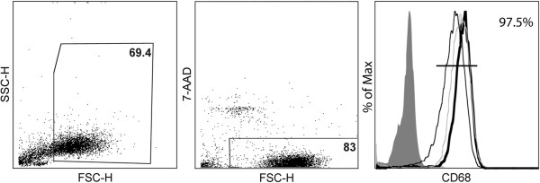
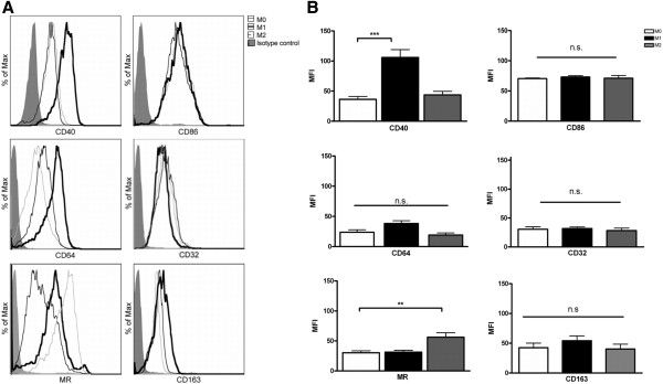
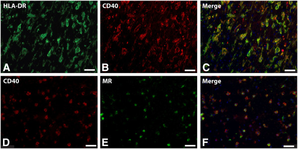
Methods: For this study, the expression of a panel of typical M1 and M2 markers on human monocyte derived M1 and M2 macrophages was analyzed using flow cytometry. This revealed that CD40 and mannose receptor (MR) were the most distinctive markers for human M1 and M2 macrophages, respectively. Using a panel of M1 and M2 markers we next examined the activation status of macrophages/microglia in MS lesions, normal appearing white matter and healthy control samples.

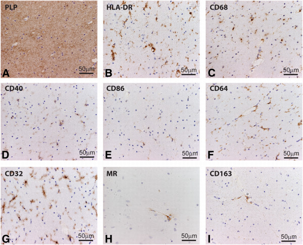
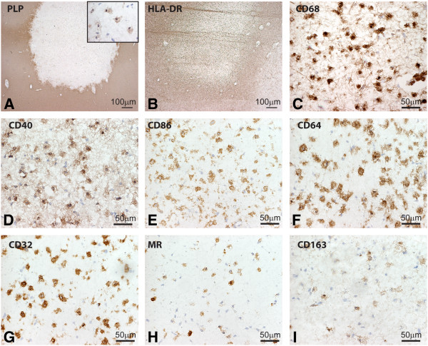
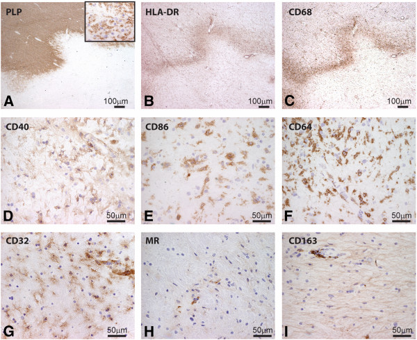
Results: Our data show that M1 markers, including CD40, CD86, CD64 and CD32 were abundantly expressed by microglia in normal appearing white matter and by activated microglia and macrophages throughout active demyelinating MS lesions. M2 markers, such as MR and CD163 were expressed by myelin-laden macrophages in active lesions and perivascular macrophages. Double staining with anti-CD40 and anti-MR revealed that approximately 70% of the CD40-positive macrophages in MS lesions also expressed MR, indicating that the majority of infiltrating macrophages and activated microglial cells display an intermediate activation status.

Conclusions: Our findings show that, although macrophages in active MS lesions predominantly display M1 characteristics, a major subset of macrophages have an intermediate activation status.


Figures

Figure 1
Figure 1
Characterization of monocyte-derived macrophages. Monocytes were isolated from peripheral blood mononuclear cells (PBMCs) of healthy donors and were cultured for 7 days in the presence of 5% normal human serum. A gate was positioned around living (7-aminoactinomycin (7-AAD) negative) cells (approximately 70%). Of the living cells approximately 97% stained positive for CD68. Representative fluorescence-activated cell sorting (FACS) plots are shown.
Figure 2
Figure 2
Expression of markers on M0, M1 and M2 macrophages stimulated in vitro. Macrophages were polarized to M1 and M2 macrophages (see materials and methods for details) and the expression of markers was analyzed by flow cytometry. In all figures the black bold line represents M1 macrophages (A). CD40 and CD64 are both shifted in mean fluorescent intensity; however, only CD40 is significantly upregulated on M1 macrophages compared to M0 macrophages. The M2 macrophage population is depicted by a grey line. Mannose receptor (MR) expression is upregulated on the M2 macrophages, whereas no differences in CD163 expression were observed comparing the different subsets. The means ± SEM were calculated from three independent experiments performed in duplicate and *P <0.05 calculated for mean fluorescent intensity (MFI) was considered significant (B).
Figure 3
Figure 3
Expression of markers for M1 and M2 phenotype in white matter of control brain. Sections of white matter of control brain were stained using immunohistochemistry. Proteolipid protein (PLP) staining shows normal abundant myelin (A) Human leukocyte antigen-DR (HLA-DR) and CD68 staining reveals positive microglia (B,C). M1 markers, including CD40, and CD86 were expressed on microglia (D,E). Antibodies directed against CD64 and CD32 clearly decorated microglia (F,G), whereas MR and CD163 were expressed by perivascular macrophages (H-I).
Figure 4
Figure 4
Expression of M1 and M2 markers in active multiple sclerosis (MS) lesions. Images were taken from the center of active demyelinating lesion. Proteolipid protein (PLP) staining shows widespread demyelination and PLP-laden macrophages (insert) (A). Intense labeling of human leukocyte antigen-DR (HLA-DR) and CD68 positive cells was observed in the center and rim of the lesion. CD68, CD40, CD86, CD64 and CD32 were markedly expressed by macrophages throughout the lesion area (D-G). Mannose receptor (MR) and CD163 were highly expressed by foamy macrophages (H,I).
Figure 5
Figure 5
CD40 and mannose receptor (MR) expression on foamy macrophages in an active multiple sclerosis (MS) lesion. Images are taken at the center of an active MS lesion stained for human leukocyte antigen-DR (HLA-DR) (A) and CD40 (B). Colocalization studies showed a clear overlap of HLA-DR and CD40 (C). Double staining with anti-CD40 (D) and -MR (E) shows that 70% of the CD40 positive cells were also MR positive, all MR positive cells are CD40 positive (F).
Figure 6
Figure 6
Expression of markers for M1 and M2 phenotype in a chronic active lesion. (A) Proteolipid protein (PLP) staining of a chronic active lesion shows massive demyelination and PLP positive macrophages in the insert. Human leukocyte antigen-DR (HLA-DR) expression was profound at the rim of chronic active lesions (B). CD68, CD40, CD86, CD64 and CD32 are all clearly expressed by microglia at the rim of the lesion. Images of macrophage markers were taken at the rim of the lesion (C-G). Mannose receptor (MR)-positive and CD163-positive macrophages were predominantly observed in the perivascular space (H-I).

References

    1. Pugliatti M, Sotgiu S, Rosati G. The worldwide prevalence of multiple sclerosis. Clin Neurol Neurosurg. 2002;104:182–191. doi: 10.1016/S0303-8467(02)00036-7. - DOI - PubMed
    1. Van der Valk P, De Groot CJ. Staging of multiple sclerosis (MS) lesions: pathology of the time frame of MS. Neuropathol Appl Neurobiol. 2000;26:2–10. doi: 10.1046/j.1365-2990.2000.00217.x. - DOI - PubMed
    1. Lassmann H. Pathology of Multiple Sclerosis, McAlpine’s MS. 3. London: Churchill Livingstone; 1998.
    1. Kigerl KA, Gensel JC, Ankeny DP, Alexander JK, Donnelly DJ, Popovich PG. Identification of two distinct macrophage subsets with divergent effects causing either neurotoxicity or regeneration in the injured mouse spinal cord. J Neurosci. 2009;29:13435–13444. doi: 10.1523/JNEUROSCI.3257-09.2009. - DOI - PMC - PubMed
    1. Abdul-Majid K-B, Stefferl A, Bourquin C, Lassmann H, Linington C, Olsson T, Kleinau S, Harris RA. Fc receptors are critical for autoimmune inflammatory damage to the central nervous system in experimental autoimmune encephalomyelitis. Scand J Immunol. 2002;55:70–81. doi: 10.1046/j.1365-3083.2002.01024.x. - DOI - PubMed

Publication types