. 2013 Mar 1:6:11.
doi: 10.1186/1756-6606-6-11.

Synaptic potentiation in the nociceptive amygdala following fear learning in mice

Affiliations

Synaptic potentiation in the nociceptive amygdala following fear learning in mice

Ayako M Watabe et al. Mol Brain. .

Abstract

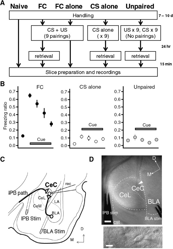
Background: Pavlovian fear conditioning is a classical form of associative learning, which depends on associative synaptic plasticity in the amygdala. Recent findings suggest that the central amygdala (CeA) plays an active role in the acquisition of fear learning. However, little is known about the synaptic properties of the CeA in fear learning. The capsular part of the central amygdala (CeC) receives direct nociceptive information from the external part of the lateral parabrachial nucleus (lPB), as well as highly processed polymodal signals from the basolateral nucleus of the amygdala (BLA). Therefore, we focused on CeC as a convergence point for polymodal BLA signals and nociceptive lPB signals, and explored the synaptic regulation of these pathways in fear conditioning.

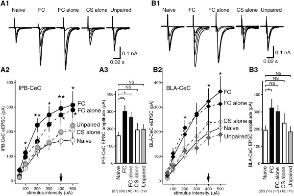
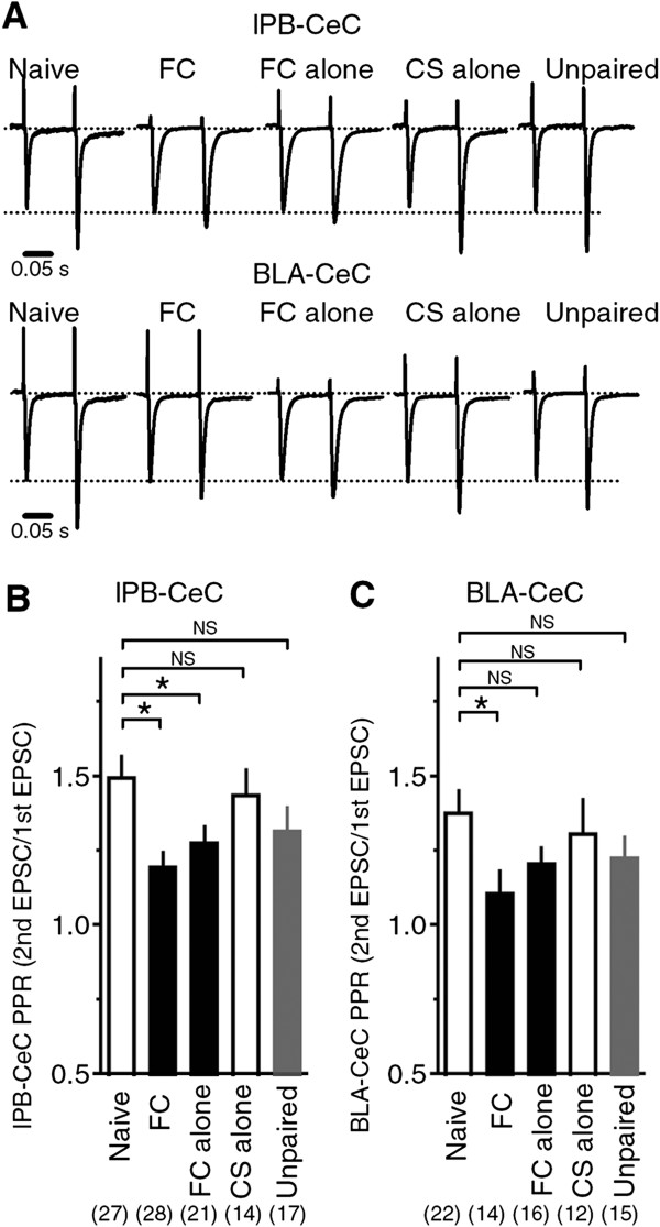
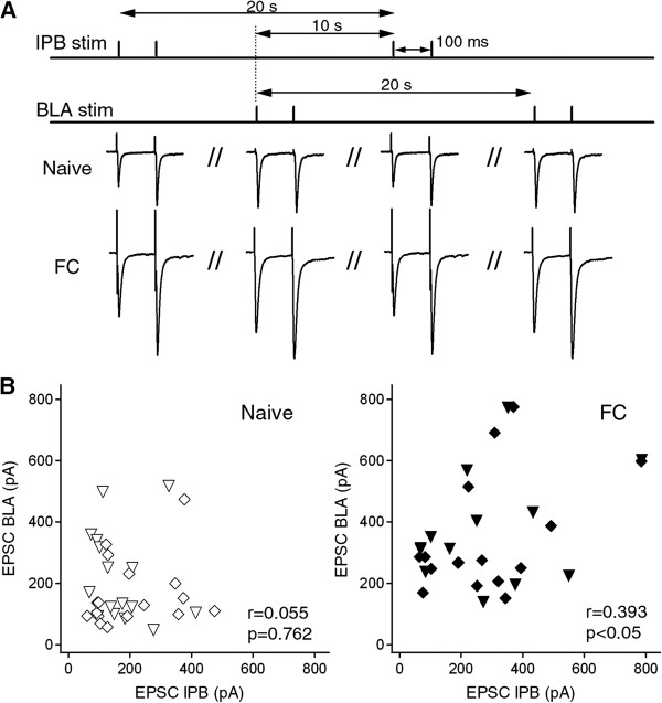
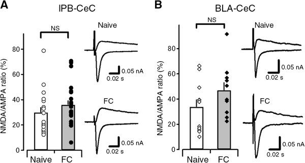
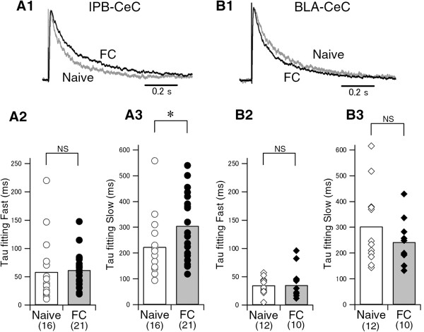
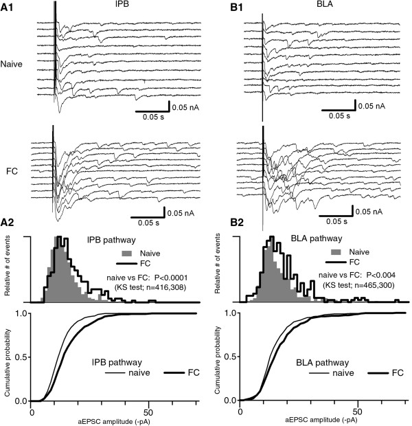
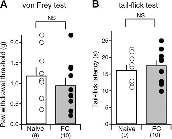
Results: In this study, we show that fear conditioning results in synaptic potentiation in both lPB-CeC and BLA-CeC synapses. This potentiation is dependent on associative fear learning, rather than on nociceptive or sensory experience, or fear memory retrieval. The synaptic weight of the lPB-CeC and BLA-CeC pathways is correlated in fear-conditioned mice, suggesting that fear learning may induce activity-dependent heterosynaptic interactions between lPB-CeC and BLA-CeC pathways. This synaptic potentiation is associated with both postsynaptic and presynaptic changes in the lPB-CeC and BLA-CeC synapses.

Conclusions: These results indicate that the CeC may provide an important locus of Pavlovian association, integrating direct nociceptive signals with polymodal sensory signals. In addition to the well-established plasticity of the lateral amygdala, the multi-step nature of this association system contributes to the highly orchestrated tuning of fear learning.


Figures

Figure 1
Figure 1
Experimental setup to examine lPB-CeC and BLA-CeC EPSCs following fear learning. A, Experimental schedules for the five different mice groups. FC, fear conditioning; CS, conditioned stimulus; US, unconditioned stimulus. B, Freezing time ratio during retrieval. The first points represent the freezing ratio during the 2-min baseline period in the chamber, while the 2nd to 5th points corresponds to 1–30 s, 31–60 s, 61–90 s and 91–120 s after the onset of the CS presentation. C, Recording configuration for lPB-CeC and BLA-CeC EPSCs. D, Oblique illumination optical images showing electrode placement (tip of a recording electrode indicated with an arrowhead) and CeC cells (bottom). Scale bars are 100 μm (top) and 10 μm (bottom).
Figure 2
Figure 2
lPB-CeC and BLA-CeC EPSCs in the five experimental groups. A1, Averaged traces of eight consecutive lPB-CeC EPSCs with increasing stimulus intensities. A2, Relationships between stimulus intensity and lPB-CeC EPSC amplitude, expressed as mean ± SEM. lPB-CeC synapse in FC mice (filled circle, solid line; n = 13–26) and FC alone mice (filled circle, dashed line; n = 18) revealed significantly enhanced synaptic transmission compared with naive mice (open circle, solid line; n = 18–27). In comparison, CS alone mice (open circle, dashed line; n = 16–18) and unpaired mice (grey circle, dashed line; n = 18–20) showed indistinguishable lPB-CeC EPSC amplitudes. *p < 0.05, **p < 0.01, analyzed with post hoc Dunnett’s t-test following ANOVA. A3, CeC EPSC amplitudes evoked by lPB stimuli of 400 μA intensity. B1, Averaged traces of eight consecutive BLA-CeC EPSCs with increasing stimulus intensities. B2, Relationships between stimulus intensity and BLA-CeC EPSC amplitude, expressed as mean ± SEM. BLA-CeC synapse in FC mice (filled diamond, solid line; n = 10–17) and FC alone mice (filled diamond, dashed line; n = 17) revealed significantly enhanced synaptic transmission compared with naive mice (open diamond, solid line; n = 18–27). On the other hand, CS alone mice (open diamond, dashed line; n = 11–14) and unpaired mice (grey diamond, dashed line; n = 15–16) showed indistinguishable BLA-CeC EPSC amplitudes. *p < 0.05, analyzed with post hoc Dunnett’s t-test following ANOVA. B3, CeC EPSC amplitudes evoked by BLA stimuli of 400 μA intensity (bottom right).
Figure 3
Figure 3
Paired-pulse ratio in lPB-CeC and BLA-CeC pathways. A, Representative traces (average of eight consecutive EPSCs) of scaled lPB-CeC EPSCs (top) and BLA-CeC EPSCs (bottom) from each group of mice. B, Averaged paired-pulse ratio (PPR) evoked by paired stimuli (100-ms interstimulus interval) in lPB-CeC synapses. Note that the PPR was significantly reduced in FC mice (n = 28) and FC alone mice (n = 21) compared with naive mice (n=27), but CS alone (n = 14) and unpaired (n = 17) mice displayed an indistinguishable PPR compared with naive mice, analyzed with post hoc Dunnett’s t-test following ANOVA. C, Averaged PPR evoked by paired stimuli (100-ms interstimulus interval) in BLA-CeC synapses. Note that the PPR was significantly reduced in FC mice (n = 28) compared with naive mice (n = 27), but FC alone (n = 21), CS alone (n = 14) and unpaired (n = 17) mice showed indistinguishable PPR compared with naive mice, analyzed with post hoc Dunnett’s t-test following ANOVA.
Figure 4
Figure 4
Correlation between lPB-CeC and BLA-CeC EPSC amplitudes. A, Experimental design for alternative stimulation of lPB and BLA pathways. B, Correlation between EPSC amplitudes of lPB-CeC (abscissa) and BLA-CeC (ordinates) synapses in naive mice (left panel, open symbols; n = 33) and fear-conditioned mice (right panel, filled symbols; n = 29). Each symbol represents data from the same set of neurons of the two pathways; the stimulation intensity of the lPB and BLA pathways was identical for these data. Different symbols represent recordings evoked by 400 μA (diamonds) and 500 μA (inverted triangles) stimulus intensities.
Figure 5
Figure 5
NMDA/AMPA ratio in lPB-CeC and BLA-CeC synapses. A, The NMDA/AMPA ratio in naive (open bar; n = 17) and fear-conditioned (filled bar; n = 22) mice at lPB-CeC synapses. Circles indicate the individual ratio for each neuron. Sample traces of NMDA current (upper trace) and AMPA current (lower trace) are superimposed for naive (top) and FC (bottom) mice. The average of 15 consecutive waves (5 min) taken 20–25 min after CNQX application, followed by changing holding potential to Vh = +40 mV, and those taken 5 min before the application of CNQX at a holding potential of Vh = −60 mV are superimposed. B, The NMDA/AMPA ratio in naive (open bar; n = 11) and fear-conditioned (filled bar; n = 20) mice in BLA-CeC synapses. Diamonds indicate individual ratio for each neuron. Sample traces of NMDA current (upper trace) and AMPA current (lower trace) are superimposed for naive (top) and FC (bottom) mice. The average of 15 consecutive waves (5 min) taken 20–25 min after CNQX application, followed by changing holding potential to Vh = +40 mV, and those taken 5 min before the application of CNQX at a holding potential of Vh = −60 mV are superimposed.
Figure 6
Figure 6
Kinetics of NMDA receptor-mediated EPSCs in lPB-CeC and BLA-CeC pathways. A1, Representative traces of scaled NMDA receptor-mediated EPSCs recorded at +40 mV in the presence of CNQX from naive (gray line) and FC (solid line) mice at lPB-CeC synapses. The averaged fast (A2) and slow (A3) decay time constants of NMDA receptor-mediated EPSCs at lPB-CeC synapses from naive (open bar) and FC (filled bar) mice. Individual values for all the cells are superimposed on the bars. B1, Representative traces of scaled NMDA receptor-mediated EPSCs recorded at +40 mV in the presence of CNQX from naive (gray line) and FC (solid line) mice at BLA-CeC synapses. Averaged fast (B2) and slow (B3) decay time constants of NMDA receptor-mediated EPSCs at BLA-CeC synapses from naive (open bar) and FC (filled bar) mice. Note that at lPB-CeC synapses, the slow decay time constant was significantly increased in FC mice compared with naive mice.
Figure 7
Figure 7
Asynchronous EPSC amplitudes in lPB-CeC and BLA-CeC synapses. A1 and B1, Representative traces using strontium solution (5 mM Sr2+, 0 mM Ca2+) in lPB-CeC (A1) and BLA-CeC (B1) synapses from naive (top panels) and FC (bottom panels) mice. A2andB2, Histograms (top panels) and cumulative plots (bottom panels) of the asynchronous EPSC (aEPSC) amplitudes in lPB-CeC (A2) and BLA-CeC (B2) synapses. Note that aEPSCs with larger amplitude emerged in FC mice so that the relative amplitude histograms were skewed towards the right. The distribution was significantly different in both lPB-CeC (p < 0.0001) and BLA-CeC (p < 0.004) synapses.
Figure 8
Figure 8
Mechanical and thermal nociceptive threshold in naive and FC mice. A, Averaged paw withdrawal threshold measured by von Frey filament test in naive (open bar) and FC (filled bar) mice. Individual values from all the mice are superimposed on each bar. B, Averaged response latencies in the tail-flick test in naive (open bar) and FC (filled bar) mice. Note that neither mechanical nor thermal nociceptive threshold differed between naive and FC mice.

References

    1. Davis M, Whalen PJ. The amygdala: vigilance and emotion. Mol Psychiatry. 2001;6(1):13–34. doi: 10.1038/sj.mp.4000812. - DOI - PubMed
    1. LeDoux JE. Emotion circuits in the brain. Annu Rev Neurosci. 2000;23:155–184. doi: 10.1146/annurev.neuro.23.1.155. - DOI - PubMed
    1. Wilensky AE, Schafe GE, Kristensen MP, LeDoux JE. Rethinking the fear circuit: the central nucleus of the amygdala is required for the acquisition, consolidation, and expression of Pavlovian fear conditioning. J Neurosci. 2006;26(48):12387–12396. doi: 10.1523/JNEUROSCI.4316-06.2006. - DOI - PMC - PubMed
    1. Zimmerman JM, Rabinak CA, McLachlan IG, Maren S. The central nucleus of the amygdala is essential for acquiring and expressing conditional fear after overtraining. Learn Mem. 2007;14(9):634–644. doi: 10.1101/lm.607207. - DOI - PMC - PubMed
    1. Busti D, Geracitano R, Whittle N, Dalezios Y, Manko M, Kaufmann W, Satzler K, Singewald N, Capogna M, Ferraguti F. Different fear states engage distinct networks within the intercalated cell clusters of the amygdala. J Neurosci. 2011;31(13):5131–5144. doi: 10.1523/JNEUROSCI.6100-10.2011. - DOI - PMC - PubMed

Publication types

Substances