Review
. 2013 Apr;34(4):243-53.
doi: 10.1016/j.tips.2013.01.009. Epub 2013 Feb 28.

Regulation of drug-induced liver injury by signal transduction pathways: critical role of mitochondria

Affiliations
Review

Regulation of drug-induced liver injury by signal transduction pathways: critical role of mitochondria

Derick Han et al. Trends Pharmacol Sci. 2013 Apr.

Abstract

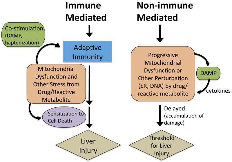
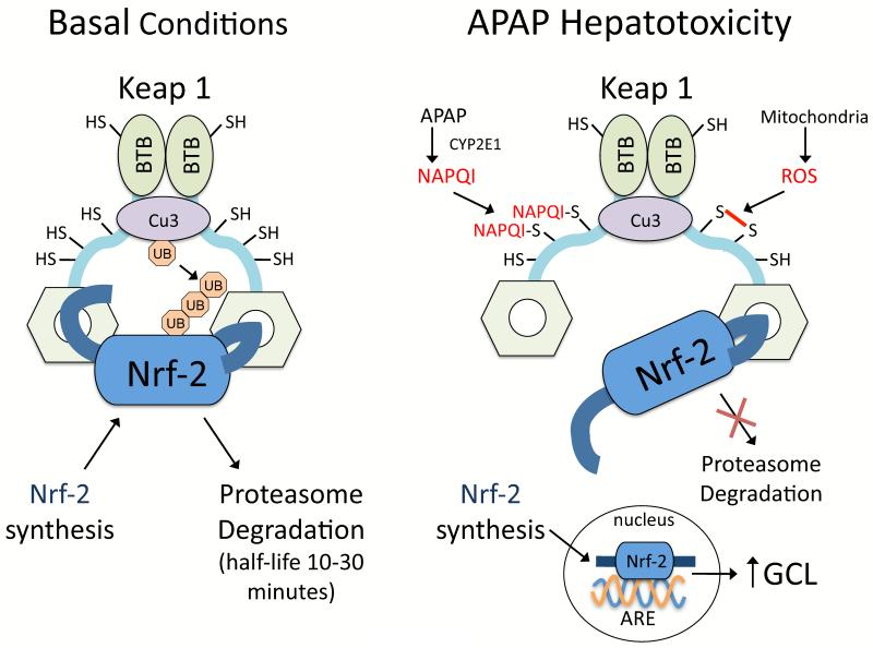
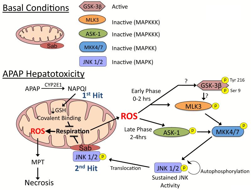
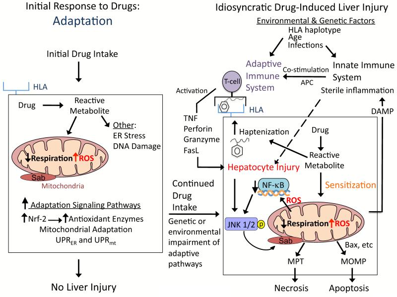
Drugs that cause liver injury often 'stress' mitochondria and activate signal transduction pathways important in determining cell survival or death. In most cases, hepatocytes adapt to the drug-induced stress by activating adaptive signaling pathways, such as mitochondrial adaptive responses and nuclear factor erythroid 2-related factor 2 (Nrf-2), a transcription factor that upregulates antioxidant defenses. Owing to adaptation, drugs alone rarely cause liver injury, with acetaminophen (APAP) being the notable exception. Drug-induced liver injury (DILI) usually involves other extrinsic factors, such as the adaptive immune system, that cause 'stressed' hepatocytes to become injured, leading to idiosyncratic DILI, the rare and unpredictable adverse drug reaction in the liver. Hepatocyte injury, due to drug and extrinsic insult, causes a second wave of signaling changes associated with adaptation, cell death, and repair. If the stress and injury reach a critical threshold, then death signaling pathways such as c-Jun N-terminal kinase (JNK) become dominant and hepatocytes enter a failsafe mode to undergo self-destruction. DILI can be seen as an active process involving recruitment of death signaling pathways that mediate cell death rather than a passive process due to overwhelming biochemical injury. In this review, we highlight the role of signal transduction pathways, which frequently involve mitochondria, in the development of DILI.


Figures

Figure 1
Figure 1. Immune-mediated and non-immune-mediated idiosyncratic DILI
Many drugs cause idiosyncratic DILI via an adaptive immune system mediated process and have strong associations with certain HLA haplotypes, even though they do not display classic characteristics of systemic immune hypersensitivity (i.e. fever, rash, eosinophilia, rapid positive rechallenge). Drugs/reactive metabolites may stress hepatocytes to stimulate the adaptive immune system. Excessive reactive metabolites bind to proteins (haptenization), and peptide-haptens exposed in the binding grove of HLA molecules then stimulate T-cells. Hepatocytes may become injured and release contents, such as mtDNA, that act as danger associated molecular pattern (DAMP) molecules that co-activate the immune system (sterile inflammation) [97]. Extensive drug-induced stress in hepatocytes may alter signaling pathways to sensitize hepatocytes to the adaptive immune system. Non-allergic DILI is believed to be mediated by biochemical injury, often involving mitochondria. The drug/reactive metabolite stress and injury to hepatocytes may need to accumulate (long latency) before manifestations of DILI are observed. DAMP molecules may also be released in this process and activate cytokines that may mediate injury. Mitochondria have a reserve respiratory capacity and significant inhibition of complexes is needed before bioenergetic capacity is impaired. Thus, drugs/reactive metabolites can damage mitochondria gradually, with no observable effects on mitochondrial respiration until a critical threshold is reached. How often non-immune idiosyncratic DILI occurs is unknown but remains plausible.
Figure 2
Figure 2. Hinge and latch model of Nrf-2 regulation by Keap1
Nrf-2 is bound to Keap1 (dimerized by binding of the BTB domains), which also has cullin-dependent E3 ubiquitin ligase complex (Cu3) attached. Cu3 ubiquitinates Nrf-2 attached to Keap1, causing Nrf-2 degradation by proteasomes. Nrf-2 bound to Keap1 consequently has a very short half-life (10-30 minutes) in hepatocytes due to continuous ubiquitination and degradation. Keap1 has critical thiols (25 cysteine residues) that act as redox and electrophile sensors. According to the “hinge and latch model”, when the thiols of Keap1 are oxidized or covalently bound by electrophillic agents such as NAPQI, a conformational change occurs in Keap1 that weakens its binding to Nrf-2. Consequently Nrf-2 becomes loosely bound to Keap1 and cannot be ubiquitinated by Cu3. Since Keap1 remains occupied by loosely attached Nrf-2 that is not degraded, newly synthesized Nrf-2 will translocate to the nucleus where it binds to the antioxidant response element (ARE) promoter on DNA important in transcribing GCL-c (catalytic component of glutamate-cysteine ligase) and other antioxidant proteins. The figure is modified from previous publications [14,16].
Figure 3
Figure 3. JNK activation during APAP-induced liver injury
A. Under basal conditions most kinases, GSK-3β being a noted exception, are inactive and reside in the cytoplasm. B. APAP hepatotoxicity involves two hits to mitochondria. Mitochondrial GSH depletion and covalent binding by NAPQI (upstream hit to mitochondria) enhance mitochondrial ROS production. ROS cause two phases of MAPK activation during APAP hepatotoxicity, an early (0-2 hours) and a late (2-4 hours) phase, which involve different initiation events. The early phase likely involves GSK-3β activating MLK3, while the late phase is mediated by ASK-1 in the liver. Both then activate MKK 4/7, which in turn activates JNK. Once activated, JNK translocates to mitochondria and binds to Sab, a scaffold protein on the outer membrane of mitochondria. JNK binding to Sab leads to sustained enhancement of mitochondrial ROS generation which has two consequences: 1) self-amplifying activation of MAPK pathways and 2) MPT and collapse of mitochondrial function. MAPK = mitogen activated protein kinase; MAPKK = MAP kinase kinase; MAPKKK – MAPK kinase kinase kinase.
Figure 4
Figure 4. Hypothesis for idiosyncratic DILI
Signaling pathways that modulate hepatocellular injury during idiosyncratic DILI probably occur in multiple waves following drug intake. The first signaling wave occurs as hepatocytes are exposed to the drug for the first time. The stress induced by the drug/reactive metabolite, in many cases due to disruption of mitochondria, causes activation of signal transduction pathways associated with adaptation to help hepatocytes cope with continuous drug intake. For most patients the activation of adaptation pathways such as Nrf-2 helps hepatocytes adjust to the drug, and no liver injury occurs. However in a minority of patients, due to genetic and environmental factors, the adaptation pathways may be overwhelmed and/or the stress imposed by continuous drug intake may cause signaling changes that sensitize hepatocytes to injury by extrinsic factors, such as the innate and/or adaptive immune system. According to the hapten model, hapten-peptides are processed and presented on HLA binding grooves of antigen-presenting cells (APC), which interact with T-cell receptors on CD4 T-cells. CD4 T-cells are activated and subsequently activate cytotoxic CD8 T-cells (CTL) that express surface FasL and release TNFα, perforin, granzyme and other cytotoxic factors. The activated CTL then target and kill the hepatocytes expressing the hapten peptide on HLA. Stressed hepatocytes may become injured because the drug/reactive metabolite inhibits pro-survival signaling pathways, such as the transcription factor, NF-κB, to sensitize hepatocytes to the immune system. If the stress and injury reach a critical threshold, then cell death signaling pathways including JNK become dominant and hepatocytes enter a failsafe mode of self-destruction. Activated JNK targets mitochondria to promote MPT and MOMP that induce apoptosis or necrosis, which manifests as DILI.
Figure 5
Figure 5. Mitochondrial adaptation to drugs
Following stress, adaptations that may occur in mitochondria include: 1) Mitochondrial unfolded protein response (UPRmt). Similar to the UPR in ER, proteins in mitochondria may become misfolded with stress and chaperone proteins may be important in proper refolding. 2) Increase in mitophagy. The rates of mitophagy (mitochondrial autophagy) are likely to increase to remove damaged or dysfunctional mitochondria (i.e. mitochondrial generating increased ROS, etc) following stress. 3) Mitochondrial remodeling. The respiratory chain of mitochondria may undergo remodeling to increase respiratory capacity to deal with metabolic stresses. 4) Mitochondrial biogenesis. Mitochondrial stress and/or injury can stimulate mitochondrial biogenesis to replace injured or damaged mitochondria, which is regulated by various proteins including PGC-1〈 and CRTC3 in the liver. 5) Changes in mitochondrial fusion-fission. Toxic and metabolic stress can cause changes in mitochondrial fusion-fission rates, which will alter mitochondrial morphology and function. APAP and other drugs cause diverse mitochondrial morphology including fragmentation, enlargement and/or spheroid formation.
Figure 6
Figure 6. Mitochondrial Sab plays a key role in TNFα-induced apoptosis
In the liver, TNFα binding to hepatocytes will activate both a cell death pathway involving JNK and caspase 8, and a survival pathway involving NF-κB activation. Following activation, NF-κB initiates transcription of antioxidant proteins (Mn-SOD, ferritin) that lower ROS levels needed to sustain JNK. TNFα-induced NF-κB activation and transcription accounts for the transient nature of JNK activation so hepatocytes are normally resistant to the cytotoxic effects of TNFα. When NF-κB is inhibited chemically or by redox alterations, JNK activity becomes sustained through a self-amplifying pathway involving JNK binding to mitochondrial Sab that enhances ROS generation. JNK also inhibits the anti-apoptotic proteins, such as cFlip and mcl-1, and activate pro-apoptotic proteins such as bax. Sustained JNK activation will then lead to MOMP and apoptosis in hepatocytes. In contrast, in APAP hepatotoxicity sustained JNK activation does not induce apoptosis because ATP depletion and redox effects inhibit caspase activation and cell death proceeds through necrosis.

References

    1. Kaplowitz N. Idiosyncratic drug hepatotoxicity. Nat Rev Drug Discov. 2005;4:489–499. - PubMed
    1. Holt M, Ju C. Drug-induced liver injury. Handb Exp Pharmacol. 2010:3–27. - PubMed
    1. Ulrich RG. Idiosyncratic toxicity: a convergence of risk factors. Annu Rev Med. 2007;58:17–34. - PubMed
    1. Han D, et al. Signal transduction pathways involved in drug-induced liver injury. Handb Exp Pharmacol. 2010:267–310. - PubMed
    1. Daly AK, et al. HLA-B*5701 genotype is a major determinant of drug-induced liver injury due to flucloxacillin. Nat Genet. 2009;41:816–819. - PubMed

Publication types

MeSH terms