. 2013 Jul;120(7):1448-53.
doi: 10.1016/j.ophtha.2012.12.009. Epub 2013 Feb 28.

Streptococcus endophthalmitis outbreak after intravitreal injection of bevacizumab: one-year outcomes and investigative results

Affiliations

Streptococcus endophthalmitis outbreak after intravitreal injection of bevacizumab: one-year outcomes and investigative results

Roger A Goldberg et al. Ophthalmology. 2013 Jul.

Abstract

Purpose: To report the 1-year clinical outcomes of an outbreak of Streptococcus endophthalmitis after intravitreal injection of bevacizumab, including visual acuity outcomes, microbiological testing, and compound pharmacy investigations by the Food and Drug Administration (FDA).

Design: Retrospective consecutive case series.

Participants: Twelve eyes of 12 patients who developed endophthalmitis after receiving intravitreal bevacizumab prepared by a single compounding pharmacy.

Methods: Medical records of patients were reviewed; phenotypic and DNA analyses were performed on microbes cultured from patients and from unused syringes. An inspection report by the FDA based on site visits to the pharmacy that prepared the bevacizumab syringes was summarized.

Main outcome measures: Visual acuity, interventions received, time to intervention, microbiological consistency, and FDA inspection findings.

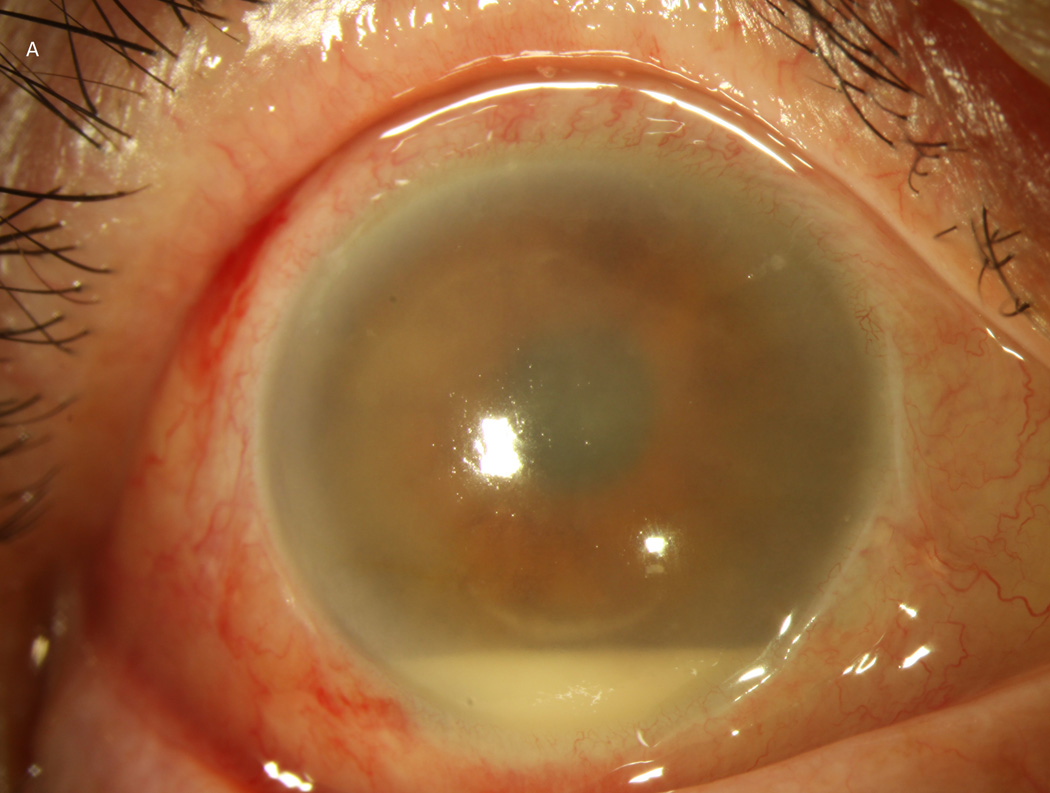
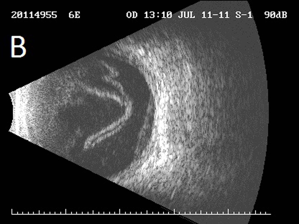
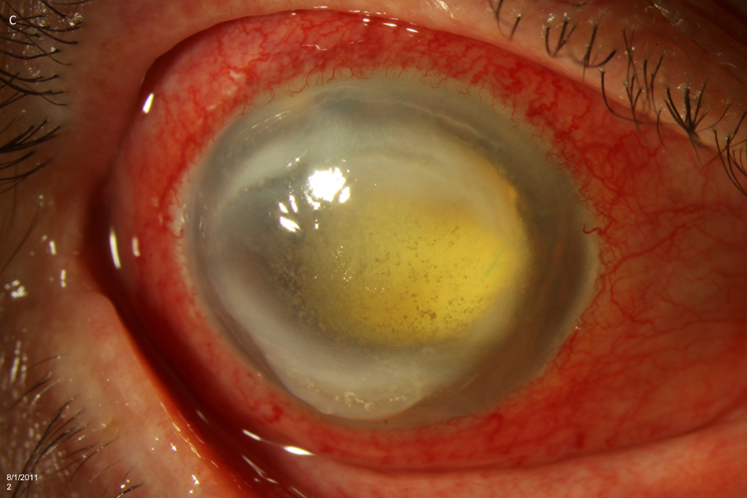
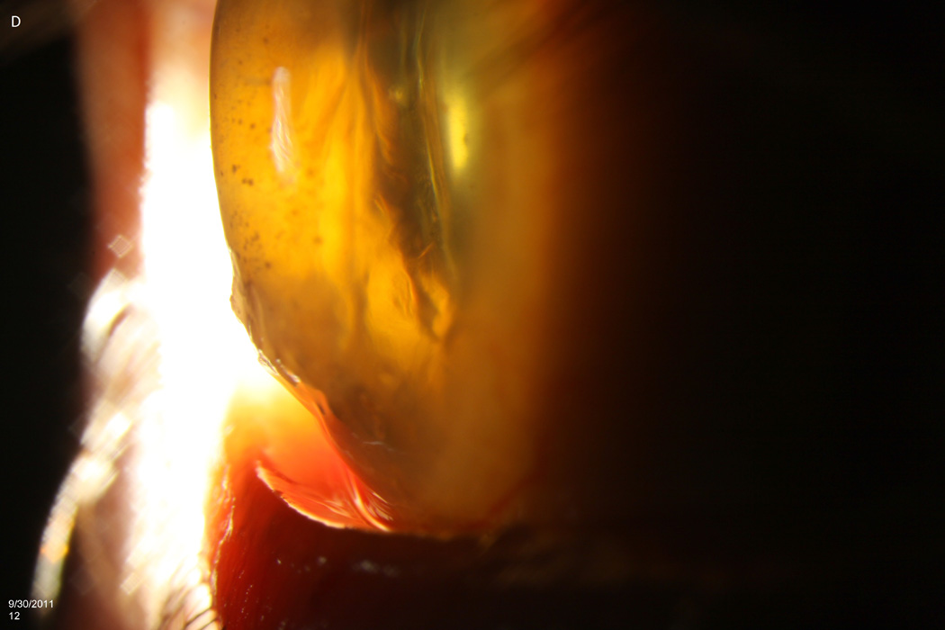
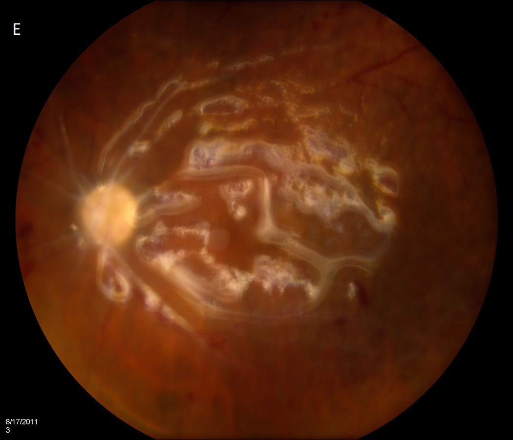
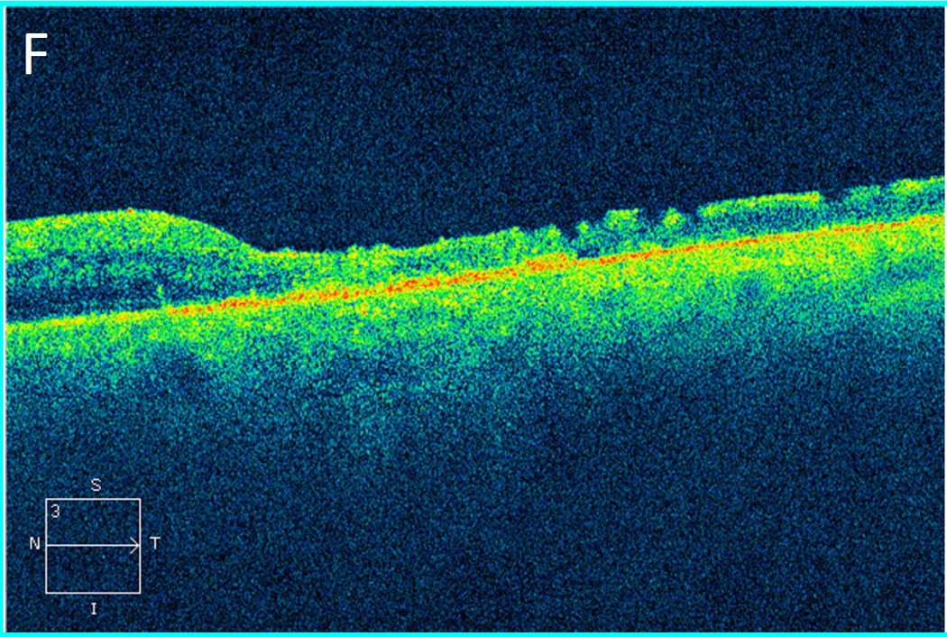
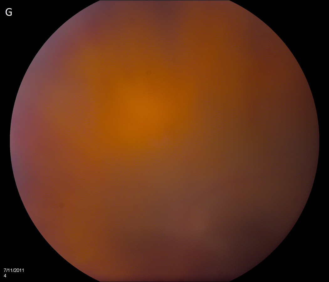
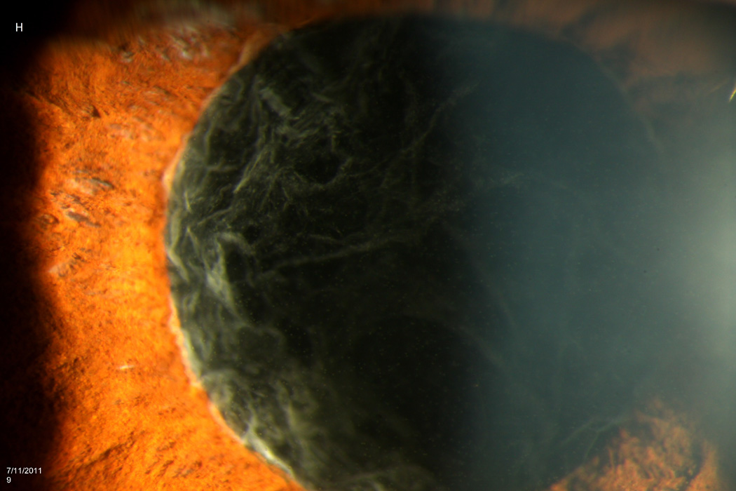
Results: Between July 5 and 8, 2011, 12 patients developed endophthalmitis after intravitreal bevacizumab from syringes prepared by a single compounding pharmacy. All patients received initial vitreous tap and injection, and 8 patients (67%) subsequently underwent pars plana vitrectomy (PPV). After 12 months follow-up, outcomes have been poor. Seven patients (58%) required evisceration or enucleation, and only 1 patient regained pre-injection visual acuity. Molecular testing using real-time polymerase chain reaction, partial sequencing of the groEL gene, and multilocus sequencing of 7 housekeeping genes confirmed the presence of a common strain of Streptococcus mitis/oralis in vitreous specimens and 7 unused syringes prepared by the compounding pharmacy at the same time. An FDA investigation of the compounding pharmacy noted deviations from standard sterile technique, inconsistent documentation, and inadequate testing of equipment required for safe preparation of medications.

Conclusions: In this outbreak of endophthalmitis, outcomes have been generally poor, and PPV did not improve visual results at 1-year follow-up. Molecular testing confirmed a common strain of S. mitis/oralis. Contamination seems to have occurred at the compounding pharmacy, where numerous problems in sterile technique were noted by public health investigators.


Conflict of interest statement

Conflicts of Interest: RAG: Emmetrope Ophthalmics. HWF: Alimera, Santen, Pfizer. DM: none. SG: none. RFI: none. PMB: none. The authors have no financial or proprietary interests in the material presented herein.

Figures

Figure 1
Figure 1
Case 2 demonstrates the marked anterior chamber reaction and a layered hypopyon (A) that most patients presented with. Echography obtained at that time confirmed the presence of a dense vitritis with prominent membranes (B). As the inflammation consolidated, several patients developed a dense ring infiltrate (C) which, in Case 8, left a prominent inferior ectasia in the cornea (D). When the view cleared sufficiently to allow for pars plana vitrectomy, the optic nerves were often pale, the vessels sclerosed, and the retina atrophic, as in Case 3 (E, F). Case 7 was the only patient to be regain her pre-injection visual acuity. She presented 3 days after her injection with milder signs and symptoms than the other patients. Her Snellen visual acuity had deteriorated from 20/40 to 20/200, with a hazy vitritis (G) and prominent pupillary fibrin (H).
Figure 1
Figure 1
Case 2 demonstrates the marked anterior chamber reaction and a layered hypopyon (A) that most patients presented with. Echography obtained at that time confirmed the presence of a dense vitritis with prominent membranes (B). As the inflammation consolidated, several patients developed a dense ring infiltrate (C) which, in Case 8, left a prominent inferior ectasia in the cornea (D). When the view cleared sufficiently to allow for pars plana vitrectomy, the optic nerves were often pale, the vessels sclerosed, and the retina atrophic, as in Case 3 (E, F). Case 7 was the only patient to be regain her pre-injection visual acuity. She presented 3 days after her injection with milder signs and symptoms than the other patients. Her Snellen visual acuity had deteriorated from 20/40 to 20/200, with a hazy vitritis (G) and prominent pupillary fibrin (H).

References

    1. McCannel CA. Meta-analysis of endophthalmitis following intravitreal injection of anti-vascular endothelial growth factor agents: causative organisms and possible prevention strategies. Retina. 2011;31:654–661. - PubMed
    1. Moshfeghi AA, Rosenfeld PJ, Flynn HW, Jr, et al. Endophthalmitis after intravitreal vascular [corrected] endothelial growth factor antagonists: a six-year experience at a university referral center. Retina. 2011;31:662–668. - PubMed
    1. CATT Research Group. Ranibizumab and bevacizumab for neovascular age-related macular degeneration. N Engl J Med. 2011;364:1897–1908. - PMC - PubMed
    1. Wen JC, McCannel CA, Mochon AB, Garner OB. Bacterial dispersal associated with speech in the setting of intravitreous injections. Arch Ophthalmol. 2011;129:1551–1554. - PubMed
    1. Schimel AM, Scott IU, Flynn HW., Jr Endophthalmitis after intravitreal injections: should the use of face masks be the standard of care? Arch Ophthalmol. 2011;129:1607–1609. - PubMed

Publication types

MeSH terms