Review
. 2013 Jul;29(7):427-37.
doi: 10.1016/j.tig.2013.01.011. Epub 2013 Feb 27.

Genetic basis of cell-cell fusion mechanisms

Affiliations
Review

Genetic basis of cell-cell fusion mechanisms

Pablo S Aguilar et al. Trends Genet. 2013 Jul.

Abstract

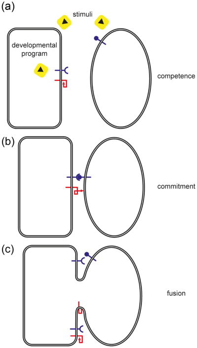
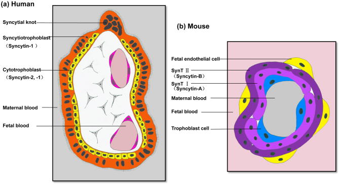
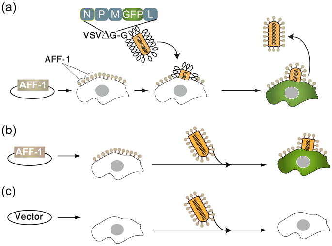
Cell-cell fusion in sexually reproducing organisms is a mechanism to merge gamete genomes and, in multicellular organisms, it is a strategy to sculpt organs, such as muscle, bone, and placenta. Moreover, this mechanism has been implicated in pathological conditions, such as infection and cancer. Studies of genetic model organisms have uncovered a unifying principle: cell fusion is a genetically programmed process. This process can be divided in three stages: competence (cell induction and differentiation); commitment (cell determination, migration, and adhesion); and cell fusion (membrane merging and cytoplasmic mixing). Recent work has led to the discovery of fusogens, which are cell fusion proteins that are necessary and sufficient to fuse cell membranes. Two unrelated families of fusogens have been discovered, one in mouse placenta and one in Caenorhabditis elegans (syncytins and F proteins, respectively). Current research aims to identify new fusogens and determine the mechanisms by which they merge membranes.


Figures

Figure 1
Figure 1. Simplified vision of steps required for cell-cell fusion
The representation of this multi-step pathway is purely schematic, and numerous cellular features are not incorporated- for example, signaling events and regulation of gene expression. A hypothetical three step scenario of heterotypic cell-cell fusion executed by a unilateral fusogen (red) is presented. (a) Competence. Differentiation into fusion-competent cells involves one or more of these complex processes: reception and response to extracellular signals, execution of developmental programs, cell polarization, cell migration, morphological changes, polarized secretion and ultimately surface display of key molecules required for the next step. (b) Commitment. This step involves cell-cell adhesion, continuous signaling and cell polarization, and consequential full exposition and/or activation of the fusogenic machinery. (c) Cell-Cell Fusion. Correct merging of plasma membranes connects both cytoplasms leading to further signaling and developmental changes. Unlike fertilization, somatic fused cells can still be competent and therefore ready for new rounds of fusion forming giant syncytia in some cases of cell-cell fusion in multicellular organisms.
Figure 2
Figure 2. Non-self fusion in S. cerevisiae and self-fusion in N. crassa is a multi-step process involving MAP kinase cell signaling
(a) Mating in S. cerevisiae involves the fusion of two haploid cells of opposite mating types (a andα) into an a/α diploid zygote. Haploid cells secrete peptide pheromones that can be detected by their cognate partners. Pheromone detection induces cell-cycle arrest, transcriptional induction of pheromone-specific genes, and polarization of growth towards the pheromone source (shmooing). Shmoo formation leads to cell-cell contact and cell wall (brown) merging. To avoid the risk of lysis caused by high internal osmotic pressure, the cell wall is degraded only at the zone of cell-cell contact. Plasma membrane fusion occurs within a few minutes after contact. Further cell wall removal allows pore expansion followed by congression and fusion of the nuclei. (b) Fusion germlings of N. crassa alternate between two physiological stages during chemotropic interaction. While two germ tubes approach each other, the cytoplasmic MAP kinase MAK-2 is recruited to the plasma membrane of the fusion tips (arrows) in an oscillating manner. Once the cells have established physical contact the kinase accumulates at the fusion point (arrowhead). Time points: time after observation started. Scale bar: 5 μm. The graph indicates fluorescence intensity of MAK2-GFP at two fusion cell tips. T1 = cell tip 1, T2 = cell tip 2 (adapted from [32]).
Figure 3
Figure 3. Podosome-like structures asymmetrically invade the myotube
(a) Stage 14 Drosophila embryo showing FCMs attached to a developing myotube (FC/myotube). Phalloidin staining reveals F-actin at the cell cortex and prominent F-actin foci at the FC/myotube//FCM interfaces. Arrowheads show late stage F-actin foci of protruding invadosomes shortly before cell-cell fusion. Scale: 2.1 μM. Images taken on a Leica SP5 63x oil immersion NA 1.4 objective [39]. (b) Schematic of (a), detailing actin focus structure in each FCM. (c) Schematic of one actin focus from (b, boxed area), indicating FCM Arp2/3 dependent pathways required for actin dynamics for the PLS and fusion. Each actin focus is surrounded by FC/myotube/FCM specific adhesion proteins (not shown) that, upon engagement, signal to actin regulators [(MyoblastCity (Mbc) ->Rac ->SCAR ->Arp2/3; BlownFuse (Blow) ->Verprolin/WIP/Solitary (Vrp1) -> WASp -> Arp2/3]. Links between these pathways exist (e.g., Blow -> Kette). Please refer to recent reviews for details on the genetic and physical interactions required for actin during myoblast fusion [–37].
Figure 4
Figure 4. Macrophage fusion is a highly-regulated, induced process
In order to fuse, macrophages acquire a fusion-competent phenotype through the integration of different signals: cytokines (e.g., RANKL + M-CSF or IL-4), cell-cell interaction (e.g., via TREM-2/DAP12 [53]), and the respective intracellular signaling pathways (NF-κB, NFATc1, STAT6, syk [52, 54, 55]). Genes essential for and upregulated during fusion include the chemokine CCL2, the putative multiple transmembrane receptors DC-STAMP and OC-STAMP, the cell adhesion molecule E-Cadherin, and the matrix metalloproteinase MMP9 [, –57]. Cytoskeletal rearrangements required for fusion are mediated by RAC1 and DOCK180 [52]. Proteinases may influence fusion by cleavage/activation/degradation of other proteins such as CD44 or Myosin IIA [58, 59]. In contrast, the matrix metalloproteinase MT1MMP seems to regulate RAC1 activity during fusion independently of proteolytic function [60]. Another crucial factor for macrophage fusion is the release of ATP via the P2X7 receptor and recognition of adenosine by the receptor Adora1 [61, 62]. In addition, exposure of phosphatidylserine and lipid recognition by CD36 as well as surface receptors recognizing macrophage-expressed ligands such as CD200 and SIRPα were shown to be involved [52]. Tetraspanins (CD9/CD81) act as molecular membrane organizers and appear to play an inhibitory role in macrophage fusion [52]. The mechanistic basis of the actual membrane fusion step has not been elucidated so far.
Figure 5
Figure 5. Comparative anatomy of the human and mouse placental syncytiotrophoblasts and the localization of Syncytins
(a) A simplified cross-section through a human chorionic villus from the first trimester placenta. It is a two-layered structure composed of a layer of mononucleated cytotrophoblast cells (yellow) and a layer of multinucleated syncytiotrophoblast (orange), which is in contact with the maternal blood. Note Syncytin-1 is expressed in both layers whereas Syncytin-2 only localizes in the cytotrophoblast cells. (b) Schematic representation of the fetal-maternal interface in the mouse placental labyrinth. The mouse placental labyrinth contains maternal and fetal blood spaces separated by three layers of trophoblast cells and a layer of fetal endothelial cells. The three layers of trophoblast cells are: a single layer of trophoblast giant cells lining the maternal blood sinusoids and two layers of syncytiotrophoblast, SynT-I and –II. SynT-II is in contact with fetal endothelial cells. Note Syncytin-A is specifically expressed in SynT-I, and Syncytin-B is only detected in SynT-II.
Figure 6
Figure 6. AFF-1 can substitute for the native fusogen glycoprotein G from Vesicular Stomatitis Virus (VSV) and fuse viral membranes to cells
(a) AFF-1 from C. elegans can complement the generation of recombinant single round infective VSVΔG-AFF-1 virus-like particles in vitro. Baby Hamster Kidney cells (BHK) expressing AFF-1 protein on the cell surface can be infected with the G-complemented VSVΔG recombinant virus (VSVΔG-G). The viral genome encodes GFP in place of the fusogenic glycoprotein G. Infection results in viral induced expression of GFP by target cells (green cytoplasm). VSVΔG-AFF-1 virus-like particles are harvested from the supernatant. (b) BHK cells can be transfected with aff-1 and infected with virus-like particles obtained in (a). The infective virus-like particles express AFF-1 on their surface instead of VSV-G-glycoprotein. (c) Cells transfected with empty vector and infected with VSVΔG-AFF-1 do not result in green cells and serve as negative controls. These experimental paradigm shows that AFF-1, in contrast to VSV-G, is necessary in both membranes to mediate virus-cell fusion [87].

References

    1. Avinoam O, Podbilewicz B. Eukaryotic cell-cell fusion families. Curr Top Membr. 2011;68:209–234. - PubMed
    1. Harrison SC. Viral membrane fusion. Nature structural & molecular biology. 2008;15:690–698. - PMC - PubMed
    1. White JM, et al. Structures and mechanisms of viral membrane fusion proteins: multiple variations on a common theme. Crit Rev Biochem Mol Biol. 2008;43:189–219. - PMC - PubMed
    1. Vaney MC, Rey FA. Class II enveloped viruses. Cell Microbiol. 2011;13:1451–1459. - PubMed
    1. Backovic M, Jardetzky TS. Class III viral membrane fusion proteins. Advances in experimental medicine and biology. 2011;714:91–101. - PubMed

Publication types

MeSH terms

LinkOut - more resources