

Influence of thickening of the inner skull table on intracranial volume measurement in older people
- PMID: 23453763
- PMCID: PMC3682185
- DOI: 10.1016/j.mri.2013.01.012
Influence of thickening of the inner skull table on intracranial volume measurement in older people
Abstract
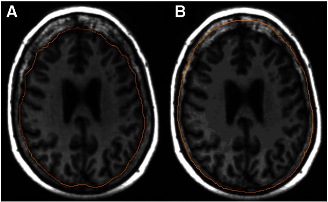
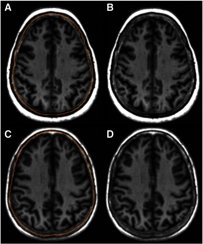
Introduction: It is generally assumed that intracranial volume (ICV) remains constant after peaking in early adulthood. Thus ICV is used as a 'proxy' for original brain size when trying to estimate brain atrophy in older people in neuroimaging studies. However, physiological changes in the skull, such as thickening of the frontal inner table, are relatively common in older age and will reduce ICV. The potential influence that inner table skull thickening may have on ICV measurement in old age has yet to be investigated.
Methods: We selected 60 (31 males, 29 females) representative older adults aged 71.1-74.3years from a community-dwelling ageing cohort, the Lothian Birth Cohort 1936. A semi-automatically derived current ICV measurement obtained from high resolution T1-weighted volume scans was compared to the estimated original ICV by excluding inner skull table thickening using expert manual image processing.
Results: Inner table skull thickening reduced ICV from an estimated original 1480.0ml to a current 1409.1ml, a median decrease of 7.3% (Z=-6.334; p<0.001), and this reduction was more prominent in women than men (median decrease 114.6 vs. 101.9ml respectively). This led to potential significant underestimations of brain atrophy in this sample by 5.3% (p<0.001) and obscured potential gender differences.
Conclusions: The effects of skull thickening are important to consider when conducting research in ageing, as they can obscure gender differences and result in underestimation of brain atrophy. Research into reliable methods of determining the estimated original ICV is required for research into brain ageing.
Copyright © 2013 Elsevier Inc. All rights reserved.
Figures

References
-
- Sahin B., Acer N., Sonmez O.F., Emirzeoglu M., Basaloglu H., Uzun A. Comparison of four methods for the estimation of intracranial volume: a gold standard study. Clin Anat. 2007;20:766–773. - PubMed
-
- Finby N., Kraft E. The aging skull: comparative Roentgen study 25 to 34 year interval. Clin Radiol. 1972;23:410–414. - PubMed
-
- Israel H. Continuing growth in the human cranial skeleton. Arch Oral Biol. 1968;13:133–137. - PubMed
-
- May H., Peled N., Dar G., Abbas J., Medlej B., Masharawi Y. Hyperostosis frontalis interna and androgen suppression. Anat Rec. 2010;239:1333–1336. - PubMed
-
- She R., Szakacs J. Hyperostosis frontalis interna: case report and review of literature. Ann Clin Lab Sci. 2004;34(2):206–208. - PubMed
Publication types
MeSH terms
Grants and funding
LinkOut - more resources
Full Text Sources
Other Literature Sources
Medical
