. 2013 Mar 28;49(6):1167-75.
doi: 10.1016/j.molcel.2013.01.035. Epub 2013 Feb 28.

AMPK-dependent degradation of TXNIP upon energy stress leads to enhanced glucose uptake via GLUT1

Affiliations

AMPK-dependent degradation of TXNIP upon energy stress leads to enhanced glucose uptake via GLUT1

Ning Wu et al. Mol Cell. .

Abstract

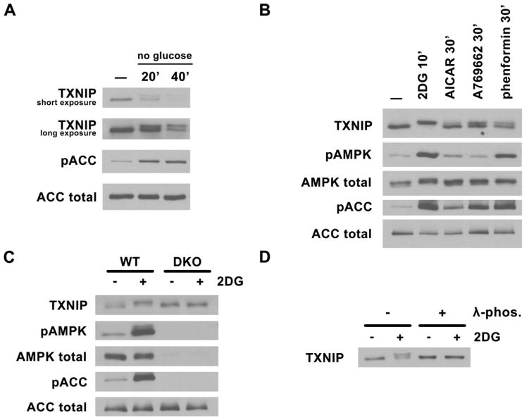
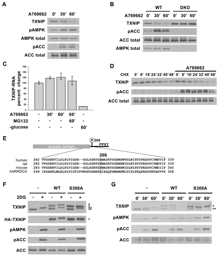
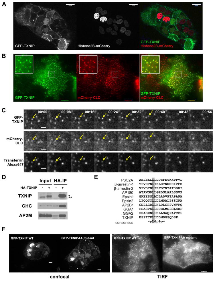
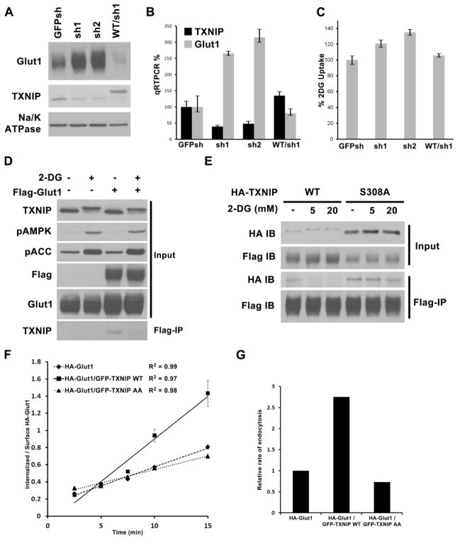
Thioredoxin-interacting protein (TXNIP) is an α-arrestin family protein that is induced in response to glucose elevation. It has been shown to provide a negative feedback loop to regulate glucose uptake into cells, though the biochemical mechanism of action has been obscure. Here, we report that TXNIP suppresses glucose uptake directly, by binding to the glucose transporter GLUT1 and inducing GLUT1 internalization through clathrin-coated pits, as well as indirectly, by reducing the level of GLUT1 messenger RNA (mRNA). In addition, we show that energy stress results in the phosphorylation of TXNIP by AMP-dependent protein kinase (AMPK), leading to its rapid degradation. This suppression of TXNIP results in an acute increase in GLUT1 function and an increase in GLUT1 mRNA (hence the total protein levels) for long-term adaptation. The glucose influx through GLUT1 restores ATP-to-ADP ratios in the short run and ultimately induces TXNIP protein production to suppress glucose uptake once energy homeostasis is reestablished.


Figures

Fig. 1
Fig. 1. AMPK-dependent TXNIP phosphorylation
Western blots of total cell lysates probed with TXNIP, phosphoAMPK (T172), total AMPK, phosphoACC (S79), and total ACC antibodies under various conditions. Activation of AMPK causes an upshift in TXNIP mobility. (A) HepG2 cells were glucose starved for 20′ and 40′ in DMEM with no glucose and 10% dialyzed FBS. (B) HepG2 cells were treated with AMPK activators: 25mM 2DG for 10′, 2mM AICAR for 60′, 1mM A769662 for 30′ and 2mM phenformin for 30′. (C) AMPK MEFs, WT and DKO (double knock-out for both α1 and α2) were treated with 25mM 2DG for 10′. (D) phosphatase treatment of endogneous TXNIP immunoprecipitated from HepG2 cells treated with 25mM 2DG abolishes the upshift.
Fig. 2
Fig. 2. AMPK phosphorylation of TXNIP on S308 accelerates its degradation
Western blots showing decreasing TXNIP levels after AMPK activation in (A) primary rat hepatocytes and (B) AMPK WT and DKO MEFs treated with 1mM A769662 for 0′, 30′ and 60′. (C) qRT-PCR analysis of TXNIP mRNA from RNA isolated from HepG2 cells after various treatments indicating short-term activation of AMPK (e.g. using A769662) does not decrease TXNIP mRNA level while glucose starvation does as reported before. The values are the average of triplicates ±STDV. (D) HepG2 cells were pretreated with cycloheximide (CHX) for 20′ to stop protein synthesis, then stimulated with A769662 to activate AMPK. Lysates harvested at indicated time points show increased rate of TXNIP protein degradation with AMPK activation. (E) Domain structure of TXNIP and multiple sequence alignment of human, rat, mouse TXNIP and human ARRDC4 around Ser308. (F) HepG2 cells stably expressing vector control, HA-WT, or HA-S308A TXNIP treated with 25mM 2DG for 10′ show that S308A mutation abolishes the phosphorylation induced protein mobility upshift after AMPK activation. (G) HepG2 cells stably expressing vector control, HA-WT or HA-S308A TXNIP were treated with 1mM A769662 for 0′, 30′ and 60′. HA-S308A TXNIP protein is degraded at a slower rate than both HA-WT and endogenous TXNIP. *: HA-tagged protein, **: endogenous protein.
Fig. 3
Fig. 3. Dual localization of TXNIP
(A) Confocal live-cell images of HepG2 cells stably expressing GFP-TXNIP and Histone2B-mCherry, show plasma membrane localization of TXNIP in addition to its nuclear localization as reported before. (B) TIRF live-cell images of HepG2 cells stably expressing GFP-TXNIP and mCherry-clathrin light chain (CLC) show some TXNIP protein localization in clathrin-coated pits (CCP). (C) HepG2 cells stably expressing GFP-TXNIP and mCherry-CLC were labeled with Alexa647-transferrin at 4°C. After rinsing off excess transferrin, time-lapse images were taken at room temperature to capture endocytosis events using a confocal microscope. For every time point, there was a 2–3 seconds delay between each fluorophore: GFP was followed by mCherry and then Alexa647. The arrow points to an endocytosed CCP that contained both GFP-TXNIP and Alexa647-transferrin. This sequence is also shown in Video S3. The scale bar is 1 μM. (D) Western blot of HA IP of lysates from mouse liver expressing adenoviral HA-TXNIP probed with clathrin heavy chain (CHC) and adaptor AP2 μ subunit (AP2M) antibodies. *: HA-tagged protein, **: endogenous protein (E) Multiple sequence alignment of di-leucine motif in various clathrin-interacting proteins. (F) Confocal and TIRF live-cell images of HepG2 cells stably expressing GFP-WT or GFP-L351AL352A TXNIP show that the LL to AA mutation abolished TXNIP localization to the CCP, but not to the plasma membrane.
Fig. 4
Fig. 4. TXNIP regulation of Glut1
Stable HepG2 cells were generated expressing control shRNA construct (GFPsh), or TXNIP knock-down shRNAs (sh1 and sh2), or knock-down cells reconstituted with HA- WT TXNIP construct that is resistant to sh1 (WT/sh1). Cells were examined for (A) Glut1 protein levels by Western blot, (B) Glut1 mRNA levels by qRT-PCR normalized to GFPsh control cells, and (C) relative rate of glucose uptake in media with 2mM glucose using trace amount of 3H-2DG, normalized to GFPsh control cells. The average values of triplicate experiments (±STDV) are reported. (D) HepG2 control cells and cells stably expressing Flag-Glut1 were treated with either water or 25mM 2DG for 10′. Flag–tag IP was carried out with the cell lysates to test interaction with endogenous TXNIP. (E) HepG2 cells stably expressing both Flag-Glut1/HA-TXNIP WT or both Flag-Glut1/HA-TXNIP S308A mutant were treated for 10′ with water, 5mM 2DG, or 20mM 2DG. Flag-tag IP was carried out to check for the effect of S308 phosphoryaltion on Glut1/TXNIP interaction. (F). HA-Glut1 endocytosis assay in TRVb-1 cells transiently transfected with HA-Glut1, HA-Glut1/GFP-TXNIP WT or HA-Glut1/GFP-TXNIP AA constructs. Cells were incubated with antibody against HA-tag on the first exofacial loop of Glut1 at 37°C and fixed after each time point. The cell surface HA-Glut1 is labeled with Cy5 secondary antibody while the endocytosed HA-Glut1 is labeled with Cy3 secondary antibody. The ratio of Cy3-to-Cy5 fluorescence signal was reported as the ratio of internalized-to-surface HA-Glut1 and plotted versus time ± SEM. R2 of the linear regression lines are shown. The data are from a representative experiment. (G). Relative endocytosis rates calculated from the slopes of linear regression from data in (F) normalized to HA-Glut1 alone control.

References

    1. Alvarez CE. On the origins of arrestin and rhodopsin. BMC evolutionary biology. 2008;8:222. - PMC - PubMed
    1. Augustin R. The protein family of glucose transport facilitators: It’s not only about glucose after all. IUBMB life. 2010;62:315–333. - PubMed
    1. Baker AF, Koh MY, Williams RR, James B, Wang H, Tate WR, Gallegos A, Von Hoff DD, Han H, Powis G. Identification of thioredoxin-interacting protein 1 as a hypoxia-inducible factor 1alpha-induced gene in pancreatic cancer. Pancreas. 2008;36:178–186. - PubMed
    1. Becuwe M, Vieira N, Lara D, Gomes-Rezende J, Soares-Cunha C, Casal M, Haguenauer-Tsapis R, Vincent O, Paiva S, Leon S. A molecular switch on an arrestin-like protein relays glucose signaling to transporter endocytosis. The Journal of cell biology. 2012;196:247–259. - PMC - PubMed
    1. Billiet L, Furman C, Cuaz-Perolin C, Paumelle R, Raymondjean M, Simmet T, Rouis M. Thioredoxin-1 and its natural inhibitor, vitamin D3 up-regulated protein 1, are differentially regulated by PPARalpha in human macrophages. Journal of molecular biology. 2008;384:564–576. - PubMed

Publication types