. 2013 Mar 18;23(6):443-52.
doi: 10.1016/j.cub.2013.02.007. Epub 2013 Feb 28.

Rheotaxis guides mammalian sperm

Affiliations

Rheotaxis guides mammalian sperm

Kiyoshi Miki et al. Curr Biol. .

Abstract

Background: In sea urchins, spermatozoan motility is altered by chemotactic peptides, giving rise to the assumption that mammalian eggs also emit chemotactic agents that guide spermatozoa through the female reproductive tract to the mature oocyte. Mammalian spermatozoa indeed undergo complex adaptations within the female (the process of capacitation) that are initiated by agents ranging from pH to progesterone, but these factors are not necessarily taxic. Currently, chemotaxis, thermotaxis, and rheotaxis have not been definitively established in mammals.

Results: Here, we show that positive rheotaxis, the ability of organisms to orient and swim against the flow of surrounding fluid, is a major taxic factor for mouse and human sperm. This flow is generated within 4 hr of sexual stimulation and coitus in female mice; prolactin-triggered oviductal fluid secretion clears the oviduct of debris, lowers viscosity, and generates the stream that guides sperm migration in the oviduct. Rheotaxic movement is demonstrated in capacitated and uncapacitated spermatozoa in low- and high-viscosity media. Finally, we show that a unique sperm motion, which we quantify using the sperm head's rolling rate, reflects sperm rotation that generates essential force for positioning the sperm in the stream. Rotation requires CatSper channels, presumably by enabling Ca(2+) influx.

Conclusions: We propose that rheotaxis is a major determinant of sperm guidance over long distances in the mammalian female reproductive tract. Coitus induces fluid flow to guide sperm in the oviduct. Sperm rheotaxis requires rotational motion during CatSper channel-dependent hyperactivated motility.


Figures

Figure 1
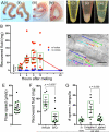
Figure 1. Increased fluid production and oviductal flow after mating
A, The mouse uterus swells after mating. (i) No fluid was recovered from the uterus at ovulation; (ii) Fluid from uteri removed immediately after mating increased by 37mg, consistent with the weight (volume) of sperm fluid; (iii, iv) Markedly increased fluid volumes in uteri 8 or 12h after mating (120, 100mg, respectively). Scale bar = 1cm. B, Accumulated uterine fluid was collected at the times indicated after mating. Fluid weights (volumes) from mated (red) and unmated (blue) females are plotted. C, Uterine fluid was collected from females (i) 20min (ii) 4h or (iii) 8h after mating, and the fluids centrifuged. D, Fluid flow in the oviduct (ampulla) 4h after mating. Migration of globular cells/debris over 3s was traced; the closed circle marks the endpoint. The central diagonal structure is a mucosal fold in the oviductal wall. Oviductal isthmus at bottom right; scale bar = 50 μm. Movie S1. E. Average globular cell migration speed =18.0 ± 1.6 μm/s at 22°C. F, Bromocriptine administration before mating inhibits fluid secretion measured 8h after mating. G, DsRed2-expressing sperm counts in the ampulla in vitro and ex vivo. Ex vivo: a portion of ampulla was dissected from females 4-5h after mating and DsRed2-expressing sperm were counted. Oviducts were removed and ligated, or left unligated, near the UTJ 60-90min after mating and then incubated at 37°C for 2-3h. The number of sperm reaching the ampulla in oviducts without an ovary averaged 31 ± 6. The increase in sperm number over the in vivo situation may result from manipulation and/or change in hormone status, perhaps detaching some sperm from the isthmal crypts and facilitating sperm migration. All graphs: mean ± s.e.m. (black bars) and median with interquartile ranges (green boxes).
Figure 2
Figure 2. Sperm rheotaxis in vitro
A, Diagram of convective flow pattern observed in warmed media. B, Laminar flow (1.5 ml/h) from a rectangular capillary at 37°C. C-F, Trajectories of mouse (C; 3s, D; 4s), and human (E-F; 5s) sperm in flow; analyzed by CASA. Scale bars: C-D; 200μm, E-F; 100μm. G, Positive rheotaxis is defined here as sperm movement against fluid flow within ± 22.5° of the forward vector of sperm movement. H, Rates of rheotaxis for mouse and human sperm (mean ± s.d.) in 5 and 3 independent experiments, respectively. Total sperm in parentheses. Movies S3, S4.
Figure 3
Figure 3. Rheotaxis of mouse sperm in low and high viscosity media
A, Capacitated (Cap) or uncapacitated (Uncap) sperm were suspended in medium with (high viscosity) or without (low viscosity) 0.3% (w/v) methylcellulose. Sperm were loaded into a capillary (at 22°C) and positive (outward flow; ~15 nl/min) or negative (inward flow; ~15 nl/min) pressure applied (37°C). The most immotile and sluggish sperm were present in the central stream (~50 μm/s flow rate), while most of motile sperm swam near the wall where the flow rate was lowest. B-C, Sperm were categorized as motile, sluggish, or immotile. Sperm movement near the end of the capillary was observed under outward (FlowOUT) or inward (FlowIN) flow in the low (B) or high (C) viscosity medium (mean ± s.d.; n = 5, total sperm counts in parentheses). Statistical significance was compared between the number of motile sperm in FlowOUT and FlowIN. P values of each experiment (compare error bars marked as a, b, c, and d) were smaller than 10-6. Movies S5-6, S8.
Figure 4
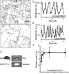
Figure 4. Sperm rotation during capacitation
A-B, Sperm trajectories in viscous solution: Sperm were incubated with 0.3% (w/v) methylcellulose-containing HEPES-buffered HTF medium for ~3min (A: uncapacitated) or 2h (B: capacitated). Scale bar: 200μm. Movie S7. C, The head of the spermatozoa is dark on phase-contrast images due to its relatively flat structure (upper panels). Sperm head light reflectance increases on phase-contrast images when the sperm heads orient vertical to the surface (lower panels). D-E, Representative results of the sperm head rolling (rotation) analysis. Uncapacitated (D) or capacitated sperm (E) incubated in HTF medium for 3h and analyzed (a.u. indicates arbitrary units). F, Increase of rotation rate during capacitation (± s.d.; ~70 sperm for each time point).
Figure 5
Figure 5. Increased sperm rotation rate in fluid flow
A, Representative uncapacitated spermatozoan turns in response to fluid flow. Images at 0.5 s intervals; black arrows mark a turning sperm. Flow direction in all panels indicated by the green arrow. Movie S9. B-C, Rotation rate of individual turning sperm; average of two 1s periods plotted at 0.5s intervals (some points overlap and are shown as single dots) (B) or sequentially over time (C). Red line in panel c indicates a turning sperm; other lines indicate sperm swimming in a straight line against fluid flow. D-E, Conditions that change sperm rotation rate. Uncapacitated sperm were incubated at 37°C and the rate of rotation measured in no flow, steady flow (14μm/s) or convergent flow (shown in (E); ± s.e.m., n = 35).
Figure 6
Figure 6. Without rotation, mouse and sea urchin sperm swim in circles
Trajectories of uncapacitated sperm from CatSper1 knockout mouse (A: 3s) or sea urchin sperm (B: 2s). Fluid flow was generated by convection or via pipette, and sperm trajectories analyzed by CASA. Scale bars: C; 200μm, D; 50μm. Green arrows show the direction of fluid flow. Movies S11-12.
Figure 7
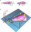
Figure 7. Model for sperm rheotaxis
A, The flagellum of an unattached sperm swimming against fluid flow was traced every 50ms and aligned by horizontal movement in the swimming direction (dotted line). Arrowhead indicates point of minimal excursion of flagellum (mef). B, Fluid flow (red arrows) re-orients sperm (yellow arrows) into the flow to reduce shear as the sperm rotate (orange arrow) and propel themselves upstream. Rotation maps out a 3-dimensional cone shape in space, which orients sperm consistently into the flow (+ rheotaxis).

References

    1. Publicover S, Harper CV, Barratt C. [Ca2+]i signalling in sperm--making the most of what you've got. Nat Cell Biol. 2007;9:235–242. - PubMed
    1. Eisenbach M, Giojalas LC. Sperm guidance in mammals - an unpaved road to the egg. Nat Rev Mol Cell Biol. 2006;7:276–285. - PubMed
    1. Fukuda N, Yomogida K, Okabe M, Touhara K. Functional characterization of a mouse testicular olfactory receptor and its role in chemosensing and in regulation of sperm motility. J Cell Sci. 2004;117:5835–5845. - PubMed
    1. Spehr M, Gisselmann G, Poplawski A, Riffell JA, Wetzel CH, Zimmer RK, Hatt H. Identification of a testicular odorant receptor mediating human sperm chemotaxis. Science. 2003;299:2054–2058. - PubMed
    1. Eisenbach M. Mammalian sperm chemotaxis and its association with capacitation. Dev Genet. 1999;25:87–94. - PubMed

Publication types