Review
. 2013 Jul 10;335(1):9-18.
doi: 10.1016/j.canlet.2013.02.036. Epub 2013 Feb 20.

Phosphatase: PP2A structural importance, regulation and its aberrant expression in cancer

Affiliations
Review

Phosphatase: PP2A structural importance, regulation and its aberrant expression in cancer

Parthasarathy Seshacharyulu et al. Cancer Lett. .

Abstract

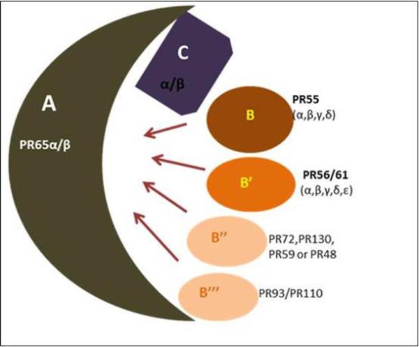
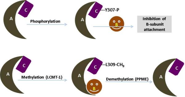
Protein Phosphatase 2A (PP2A) is an important and ubiquitously expressed serine threonine phosphatase and regulates the function by dephosphorylating many critical cellular molecules like Akt, p53, c-Myc and β-catenin. It plays a critical role in cellular processes, such as cell proliferation, signal transduction and apoptosis. Structurally, it is multifarious as it is composed of catalytic, scaffold and regulatory subunits. The catalytic and scaffold subunits have two isoforms and the regulatory subunit has four different families containing different isoforms. The regulatory subunit is the most diverse with temporal and spatial specificity. PP2A undergoes post-translational modifications (i.e. phosphorylation and methylation), which in turn, regulates its enzymatic activity. Aberrant expression, mutations and somatic alterations of the PP2A scaffold and regulatory subunits have been observed in various human malignancies, including lung, breast, skin and colon cancer, highlighting its role as a 'tumor suppressor'. This review is focused on the structural complexity of serine/threonine phosphatase PP2A and summarizes its expression pattern in cancer. Additionally, the PP2A interacting and regulatory proteins and substrates are also discussed. Finally, the mouse models developed to understand the biological role of PP2A subunits in an in vivo model system are also reviewed in this article.


Figures

Figure 1
Figure 1. Schematic representation of the structural diversity of the PP2A holoenzyme complex
PP2A enzymes are heterotrimers consisting of core dimer sscaffold (A) and a ccatalytic (C) subunit that is associated with one of the rregulatory (B) subunits. The scaffold A and catalytic C are encoded by two distinct genes α and β. The α and β isoforms of the catalytic subunit share 97% homology. However, in the typical cell, the catalytic α isoform predominates which is 10 times more abundant than the β isoform. Similar to catalytic subunit α and β isoforms of the scaffold subunit also share 86% identity in their primary sequence. The B-type subunits were further categorized into four unrelated families: B (PR55), B’ (PR56/61), B” (PR72/130) and B’” (PR93/110). Within each B-type family, distinct genes encode various structurally related isoforms such as α, β, γ and δ.
Figure 2
Figure 2. Post transitional modification of PP2A
PP2A is being regulated by post translation modification, auto-regulation, subunit diversity and substrate protein interaction. Phosphorylation and methylation are two major modifications that have been shown to modulate PP2A subunit associations and catalytic efficiency. Upon reversible phosphorylation of tyrosine residue 307 (located in the C-terminal part of PP2A catalytic subunit) by receptor associated tyrosine kinases (pp60v-src, pp56lck, EGF and insulin receptors) results in decreased phosphatase activity of PP2A. In addition to tyrosine phosphorylation, PP2A can also be auto-phophorylated on threonine residues by activated protein kinases. Apart from the PP2A catalytic subunit, the regulatory subunits are also subjected to phosphorylation. Notably, PR61δ by PKA and PR 61α by PKR are the best evidence for the change in the PP2A activity. Similarly, methylation of PP2A occurs at leucine 309 residue by PP2A-methyltransferase and enhances the binding affinity of the AC core dimer toward a distinct regulatory subunit.
Figure 3
Figure 3. Representation of PP2A regulation and signaling pathways
Methylation and phosphorylation are two major post translation modifications of PP2A. The conserved sequence TPDYFL in PP2Ac undergoes phosphorylation and methylation, respectively. Phosphorylation of Y307 by receptor associated tyrosine kinases effectively decreases the PP2A activity by inhibiting the interaction of the catalytic subunit with the scaffold subunit. On the other hand, the addition of the methyl group to PP2Ac by LCMT1 at L309 by PP2A-methyltransferase (PPMT) will enhance the specific binding of the AC dimer to the distinct B regulatory subunit providing the enzymatic activity. PP2A activity is indispensable for every cell and takes part in the majority of the cellular pathways. Phosphorylation at Thr-308 and Ser-473 leads to activation of Akt and is associated with PP2A activation. PP2A also possesses a vital role in the Wnt signaling pathway and phosphorylation of PP2A-B56 directs the activation of the Wnt pathway. In addition to these pathways, PP2A activity is indispensable in apoptosis. BAD (pro-apoptotic) and Bcl-2 are also regulated by PP2A. Phosphorylation of BAD suppresses, and its dephosphorylation by PP2A promotes pro-apoptotic activity. In addition, phosphorylation of Bcl-2 activates, and its dephosphorylation by PP2A suppresses anti-apoptotic activity. Thus, this diagrammatic representation will elucidate the importance of PP2A during cell growth survival and apoptosis.

References

    1. Mumby MC, Walter G. Protein serine/threonine phosphatases: structure, regulation, and functions in cell growth. Physiol Rev. 1993;73:673–699. - PubMed
    1. Johnson SA, Hunter T. Kinomics: methods for deciphering the kinome. Nat. Methods. 2005;2:17–25. - PubMed
    1. Alonso A, Sasin J, Bottini N, Friedberg I, Friedberg I, Osterman A, Godzik A, Hunter T, Dixon J, Mustelin T. Protein tyrosine phosphatases in the human genome. Cell. 2004;117:699–711. - PubMed
    1. Tonks NK. Protein tyrosine phosphatases: from genes, to function, to disease. Nat. Rev. Mol. Cell Biol. 2006;7:833–846. - PubMed
    1. Olsen JV, Blagoev B, Gnad F, Macek B, Kumar C, Mortensen P, Mann M. Global, in vivo, and site-specific phosphorylation dynamics in signaling networks. Cell. 2006;127:635–648. - PubMed

Publication types

MeSH terms