. 2013 Apr 1;12(7):1083-90.
doi: 10.4161/cc.24090. Epub 2013 Mar 1.

Chk1-Mad2 interaction: a crosslink between the DNA damage checkpoint and the mitotic spindle checkpoint

Affiliations

Chk1-Mad2 interaction: a crosslink between the DNA damage checkpoint and the mitotic spindle checkpoint

Rosaria Chilà et al. Cell Cycle. .

Abstract

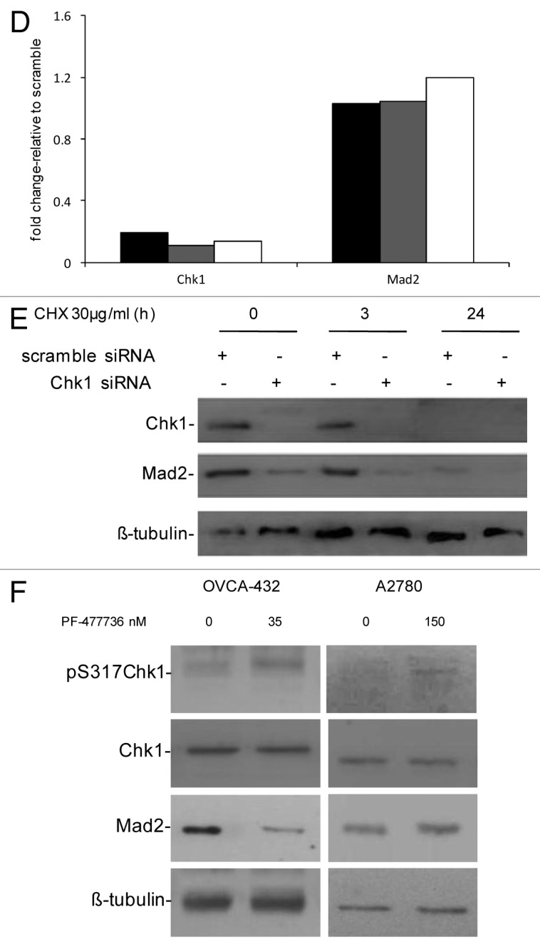
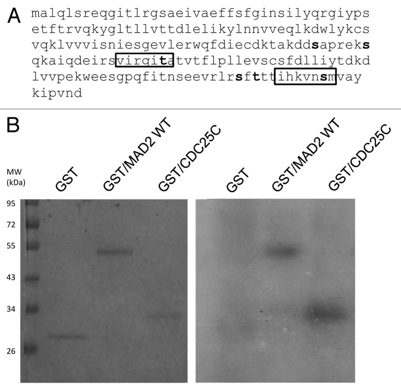
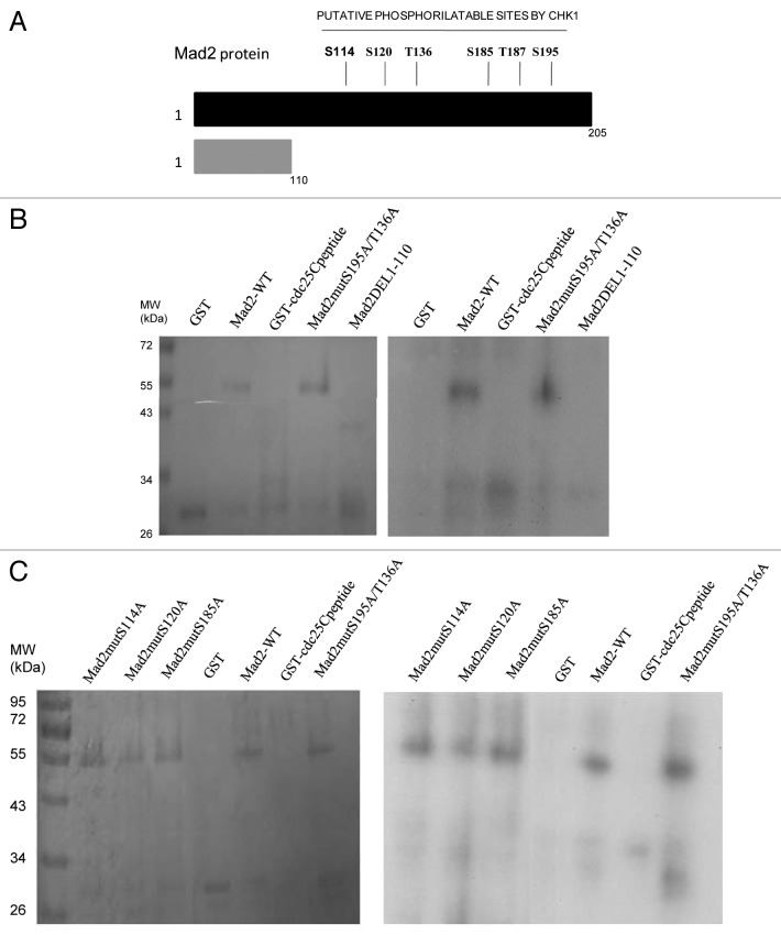
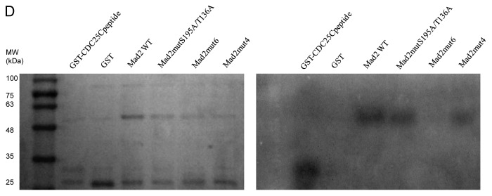
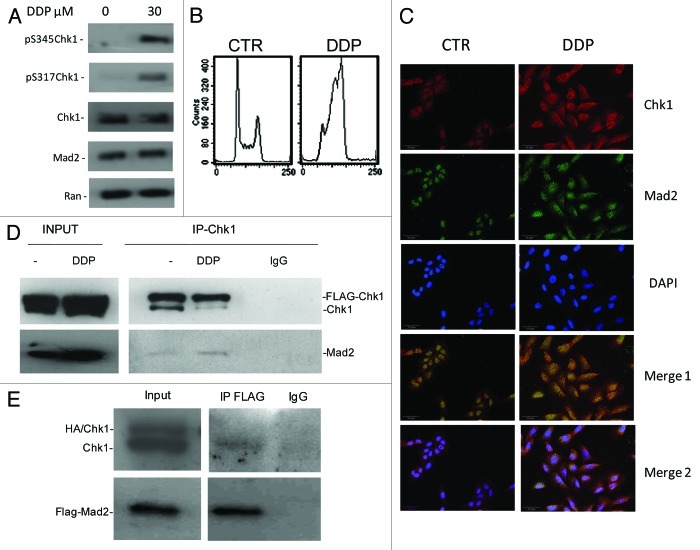
Chk1 is implicated in several checkpoints of the cell cycle acting as a key player in the signal transduction pathway activated in response to DNA damage and crucial for the maintenance of genomic stability. Chk1 also plays a role in the mitotic spindle checkpoint, which ensures the fidelity of mitotic segregation during mitosis, preventing chromosomal instability and aneuploidy. Mad2 is one of the main mitotic checkpoint components and also exerts a role in the cellular response to DNA damage. To investigate a possible crosslink existing between Chk1 and Mad2, we studied Mad2 protein levels after Chk1 inhibition either by specific siRNAs or by a specific and selective Chk1 inhibitor (PF-00477736), and we found that after Chk1 inhibition, Mad2 protein levels decrease only in tumor cells sensitive to Chk1 depletion. We then mapped six Chk1's phosphorylatable sites on Mad2 protein, and found that Chk1 is able to phosphorylate Mad2 in vitro on more than one site, while it is incapable of phoshorylating the Mad2 form mutated on all six phosphorylatable sites. Moreover our studies demonstrate that Chk1 co-localizes and physically associates with Mad2 in cells both under unstressed conditions and after DNA damage, thus providing new and interesting evidence on Chk1 and Mad2 crosstalk in the DNA damage checkpoint and in the mitotic spindle checkpoint.

Keywords: Chk1; DNA damage checkpoint; Mad2; mitotic spindle checkpoint.


Figures

None
Figure 1. (A) Western blot analysis of Chk1, Mad2 and Actin in U2OS, OVCAR8, OVCA432, A2780 and OVCAR-5 cells 72 h after either scramble or Chk1 siRNA transfection. (B) Densitometric analysis of Mad2 protein levels in the cell lines 72 h after siRNA Chk1 transfection, compared with the siRNA scramble transfected cells. Data are percentages of siRNA scramble controls, representing the ratio of Mad2 to actin. (C) Schematic representation of the percentage of cells surviving at 72 h after transfection with siRNA Chk1. Data are expressed as the percentages of scramble transfected cells and are the mean ± SD of two independent experiments.
None
Figure 1D–F. (D) Chk1 and Mad2 mRNA expression in U2OS (black bars), OVCAR-8 (gray bars) and OVCA-432 (white bars) by real-time PCR 72 h after transfection with siRNA Chk1. Chk1 and Mad2 expression levels are normalized to the internal mRNA levels of actin and are represented as the fold change from scramble transfected samples. (E) Western blot analysis of Chk1, Mad2 and β-tubulin in U2OS cells transfected with either scramble or Chk1 siRNA and 72 h post-transfection either untreated or treated with CHX 30 µg/ml for 3 and 24 h. (F) Western blot analysis of Mad2, pS317-Chk1, Chk1 and β-tubulin in OVCA-432 and A2780 cells 72 h after treatment with 35 or 150 nM of the Chk1 inhibitor PF-00477736 compared with untreated cells.
None
Figure 2. (A) Schematic representation of the Chk1 phosphorylatable sites on the Mad2 protein sequence. Specific S/T sites are evidenced in bold type. The boxes illustrate the two complete Chk1 phosphorylatable sites of Mad2. (B) In vitro kinase assay using wt recombinant GST-Chk1 in the presence of 32γ ATP and the positive recombinant substrate GST-Cdc25C, the putative recombinant substrate GST-Mad2 and the negative control GST. Left: SDS-PAGE stained with comassie blue. Right: autoradiography.
None
Figure 3. A Schematic representation of the Mad2 protein region with Chk1 phosphorylatable sites and the C-terminally truncated part with no phosphorylatable sites (1–110). (B–D) In vitro kinase assay using wt recombinant GST-Chk1 in the presence of 32γ ATP and the positive recombinant substrate GST-Cdc25C, the putative recombinant substrate GST-Mad2 wt, the mutated proteins S195A/T136A and the DEL1–110 (B), the mutated proteins S114A, S120A and S185A (C) and the mutated form in 4 and 6 phosphorylatable sites (D). GST, negative control. Left: SDS-PAGE stained with comassie blue. Right: autoradiography.
None
Figure 3D. See Figure 3 legend.
None
Figure 4. (A) Western blot analysis of pS345-Chk1, pS317-Chk1, Chk1, Mad2 and Ran in U2OS cells 24 h after treatment with 30 µM of DDP and in untreated cells. (B) Analysis of DNA content by FACS 24 h after the end of treatment with 30 µM of DDP and in untreated samples. (C) Chk1, Mad2 and DAPI staining in U2OS cells. Merge 1: Mad2 and Chk1 overlay. Merge 2: Chk1, Mad2 and DAPI overlay. (D) Exogenous (Flag-Chk1) and endogenous Chk1 immunoprecipitation from cell extracts of U2OS overexpressing Flag-Chk1 and Mad2, either untreated or 24 h after treatment with 30 µM DDP using a rabbit polyclonal Chk1 antibody. Immunoprecipitation with Rabbit IgG: negative control of the experiment. Immunoprecipitates probed by western blot analysis for Chk1 and Mad2 protein levels. Inputs: total cell extracts, either untreated or treated with DDP (the experiment showed is representative of two independent experiments). (E) Exogenous Flag-Mad2 immunoprecipitation from cell extracts of U2OS overexpressing Flag-Mad2 and HA-Chk1, using a mouse monoclonal Flag antibody. Immunoprecipitation with Mouse IgG: negative control of the experiment. Immunoprecipitates probed by western blot analysis for Chk1 and Mad2 protein levels. Inputs: total cell extracts.

References

    1. Suijkerbuijk SJ, Kops GJ. Preventing aneuploidy: the contribution of mitotic checkpoint proteins. Biochim Biophys Acta. 2008;1786:24–31. - PubMed
    1. Draviam VM, Xie S, Sorger PK. Chromosome segregation and genomic stability. Curr Opin Genet Dev. 2004;14:120–5. doi: 10.1016/j.gde.2004.02.007. - DOI - PubMed
    1. Laiho M, Latonen L. Cell cycle control, DNA damage checkpoints and cancer. Ann Med. 2003;35:391–7. doi: 10.1080/07853890310014605. - DOI - PubMed
    1. Fang G, Yu H, Kirschner MW. The checkpoint protein MAD2 and the mitotic regulator CDC20 form a ternary complex with the anaphase-promoting complex to control anaphase initiation. Genes Dev. 1998;12:1871–83. doi: 10.1101/gad.12.12.1871. - DOI - PMC - PubMed
    1. Wassmann K, Benezra R. Mad2 transiently associates with an APC/p55Cdc complex during mitosis. Proc Natl Acad Sci USA. 1998;95:11193–8. doi: 10.1073/pnas.95.19.11193. - DOI - PMC - PubMed

MeSH terms