. 2013 Apr;20(4):469-76.
doi: 10.1038/nsmb.2519. Epub 2013 Mar 3.

SOCS3 binds specific receptor-JAK complexes to control cytokine signaling by direct kinase inhibition

Affiliations

SOCS3 binds specific receptor-JAK complexes to control cytokine signaling by direct kinase inhibition

Nadia J Kershaw et al. Nat Struct Mol Biol. 2013 Apr.

Abstract

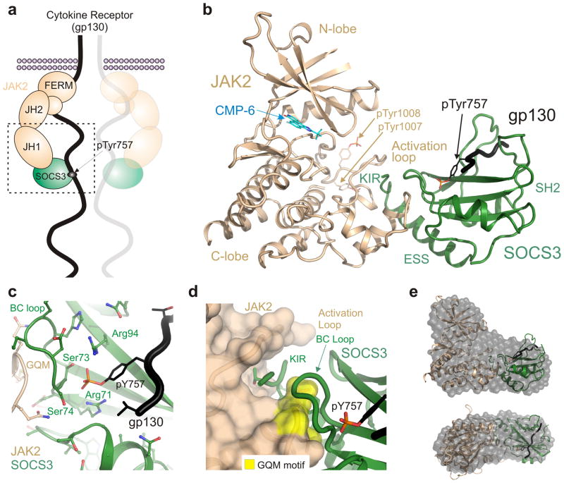
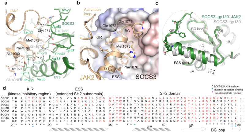
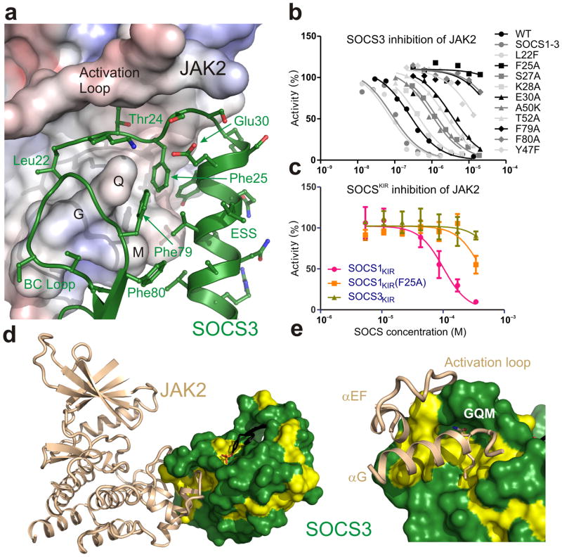
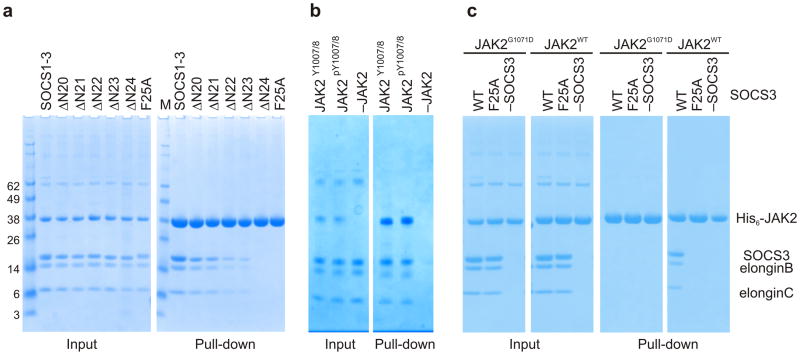
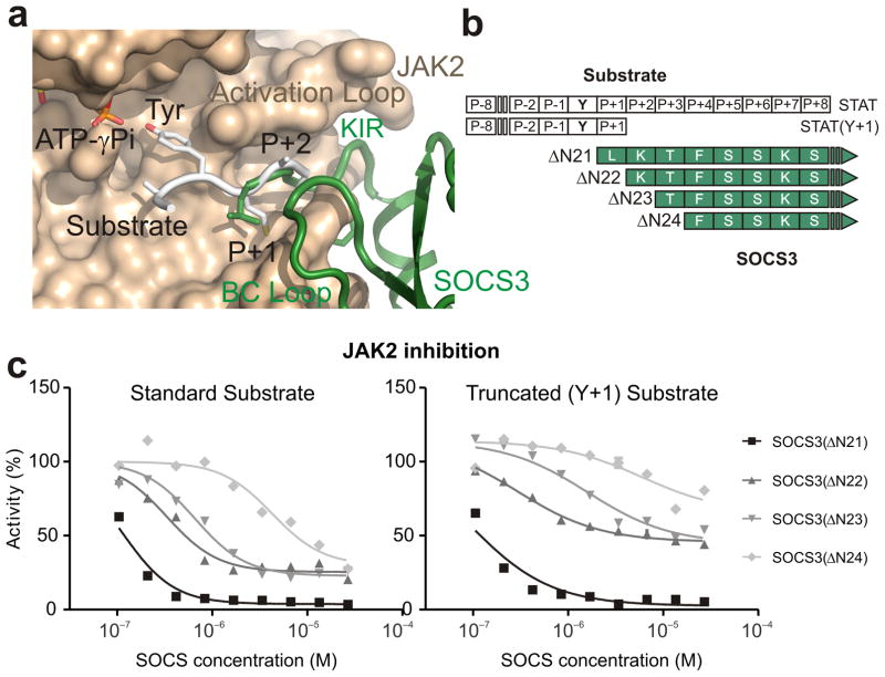
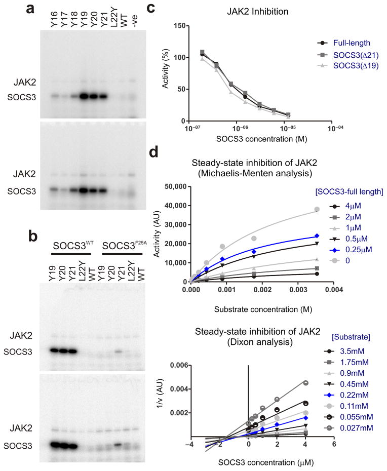
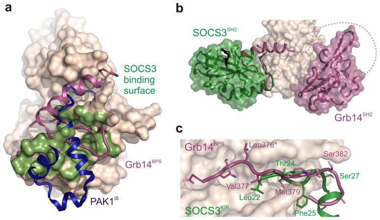
The inhibitory protein SOCS3 plays a key part in the immune and hematopoietic systems by regulating signaling induced by specific cytokines. SOCS3 functions by inhibiting the catalytic activity of Janus kinases (JAKs) that initiate signaling within the cell. We determined the crystal structure of a ternary complex between mouse SOCS3, JAK2 (kinase domain) and a fragment of the interleukin-6 receptor β-chain. The structure shows that SOCS3 binds JAK2 and receptor simultaneously, using two opposing surfaces. While the phosphotyrosine-binding groove on the SOCS3 SH2 domain is occupied by receptor, JAK2 binds in a phosphoindependent manner to a noncanonical surface. The kinase-inhibitory region of SOCS3 occludes the substrate-binding groove on JAK2, and biochemical studies show that it blocks substrate association. These studies reveal that SOCS3 targets specific JAK-cytokine receptor pairs and explains the mechanism and specificity of SOCS action.


Figures

Figure 1
Figure 1. The structure of a JAK/SOCS/gp130 complex and the two interfaces
(a) Schematic of SOCS3 inhibiting JAK2 whilst bound to gp130 (shared receptor for IL-6, LIF etc). JAK2 consists of four distinct domains and the C-terminal kinase domain (JH1) interacts with SOCS3. The boxed region indicates the ternary complex structure solved in this work (b) Ribbon diagram of the JAK2 (beige)/SOCS3 (green)/gp130 (black) complex. The gp130 peptide is located in the canonical pTyr-binding groove on the SH2 domain of SOCS3 whilst the opposing face of the SH2 domain contacts JAK2. (c) SOCS3 binds gp130 via the canonical pTyr-binding groove on the SH2 domain with the BC loop (S73-F79) of SOCS3 contacting both JAK2 and gp130 via opposing faces. The co-ordination of the phosphate moiety from gp130 pY757 is identical to that seen in the absence of JAK2. (d) The short KIR motif of SOCS3 sits in a groove bordered by the activation loop and GQM motif (yellow) of JAK2. (e) The molecular envelope of the JAK2/SOCS3/gp130 complex in solution calculated from SAXS data by performing 10 independent DAMMIF ab initio bead reconstructions (grey spheres) superimposed with the complex crystal structure (as shown in panel a). Upper panel shown in the same orientation as panel b; lower panel, top view. Data collection statistics and validation are shown as Supplementary materials.
Figure 2
Figure 2. The SOCS3/JAK2 interaction
(a) The hydrophobic SOCS3/JAK2 interface. Important residues are labeled and a selection of van der Waals contacts shown as dotted lines. Color scheme is the same as Figure 1. (b) Residues from the GQM motif and αG helix of JAK2 bind a concave hydrophobic surface on SOCS3 formed by the KIR, BC loop and extended SH2 subdomain (ESS). JAK2 is shown in ribbon representation and SOCS3 as an electrostatic surface (+/− 250mV). The GQM motif and Phe1076 from αG of JAK2 are highlighted. (c) Comparison of the SOCS3 structure in isolation (PDB:2HMH, white) and in complex with JAK2 (this work, green). The ESS helix can be seen to undergo a translation of half a helical turn upon binding JAK2 whilst the KIR (which is unstructured in the absence of JAK2) adopts an extended structure. The orientation of SOCS3 is the same as in (b). (d) Sequence conservation of the JAK2-binding interaction surface between SOCS1 and SOCS3. Conserved residues are shown in red.
Figure 3
Figure 3. The JAK2 binding epitope is composed of the KIR, ESS and SH2 domain and is conserved between SOCS3 and SOCS1
(a) Cut-away view of the JAK2-binding epitope on SOCS3. SOCS3 is shown as a ribbon diagram and JAK2 as an electrostatic surface (+/− 250mV). The SOCS3 KIR (Leu22–Ser29), which is unstructured in isolation, folds back underneath the BC loop and sits in a groove formed by the JAK2 activation loop and GQM motif (labeled). SOCS3 Phe25 sits in a deep hydrophobic pocket formed by residues from both SOCS3 and JAK2. The interface is mostly hydrophobic. (b) IC50 plots of SOCS3 point mutants. These assays highlight SOCS3 Phe25, Phe79 and Phe80 as being required for inhibition. The curves shown are an average of duplicate experiments. (c) An isolated KIR peptide from SOCS1 inhibits JAK2 with a 0.1mM IC50. Error bars are SD from triplicate experiments. (d) SOCS3/JAK2 interface highlights the conservation of residues in the JAK2-binding epitope. Conserved residues between SOCS3 and SOCS1 shown in yellow. (e) Close-up of the interface shown in (d). Color scheme as in (d).
Figure 4
Figure 4. The KIR is required for JAK binding
(a) Co-precipitation experiments show that SOCS3 binds JAKJH1 provided the kinase inhibitory region is intact. Lanes 2–6 show there is a gradual loss of binding as the N-terminal residues of the SOCS3 KIR are removed, as well as when Phe25 is mutated to Alanine. The SOCS1-SOCS3 chimera is shown as a positive control (b) Co-precipitation experiments show that SOCS3binds activated (pY1007/8) and dephosphorylated (Y1007/8) JAK2JH1 with similar affinity. Dephosphorylated JAK2JH1 was prepared by co-expressing it with the phosphatase PTP1B and then used in co-precipitation experiments with SOCS3 as described in panel a. A western blot of this experiment probed with a pY1007/8 specific antibody shows that JAK2 co-expressed with PTB1B was >95% dephosphorylated (Supplemental Figure 5) (c) Co-precipitation experiments show that SOCS3 does not bind JAK2JH1 if the JAK2 GQM motif is mutated (G1071D). SOCS3F25A is shown as a negative control.
Figure 5
Figure 5. SOCS3 inhibits JAK2 by blocking substrate binding
(a) Model of a substrate peptide (white) bound to JAK2 based on the IRK–substrate–ATP structure (PDB 1IR3) shows that the KIR of SOCS3 would block substrate binding. L22 is located where the P+1 residue would reside (both are shown in stick representation). (b) Schematic of the predicted overlap with substrate binding for the constructs and substrates used in these experiments. (c) Kinase inhibition assays performed with constructs of SOCS3 truncated at the N-terminal end of the KIR show that constructs lacking 1–3 residues only partially inhibit JAK2 activity. Left, kinase inhibition experiments using the standard STAT5b peptide (Y+8) as a substrate. Right, as for left but using a C-terminally truncated (Y+1) substrate. Inhibition of STAT5bY+1 by SOCS3ΔN22,23 is only 50% complete under saturating conditions.
Figure 6
Figure 6. Residues upstream of the SOCS3 KIR act as a pseudosubstrate
(a) Radioactive kinase assays show that constructs of SOCS3 with a tyrosine 1–3 residues upstream of the KIR are good substrates for JAK2. Reactions were performed for 1 minute (upper panels) and 2 minutes (lower panels) and then analyzed via SDS-PAGE followed by autoradiography. Experiments performed in the absence of SOCS3 (−ve) are shown as a control. (b) As for (a) but SOCS3 F25A mutants were used as controls to show that tyrosines upstream of the KIR are only good substrates for JAK2 when forced into close proximity of the active site by the remainder of SOCS3. (c) Full-length SOCS3 inhibits JAK2 with an identical IC50 to SOCS3 lacking the first 19 or 21 residues. (d) Steady-state inhibition of JAK2 by SOCS3 gives non-competitive inhibition as analyzed by Michaelis-Menten (upper) and Dixon (lower) plots. The average of duplicate experiments are shown in each case.
Figure 7
Figure 7. Comparison of three kinase inhibitors: SOCS3, PAK1IS and Grb14
(a) SOCS3 binds to a surface on the JAK2 kinase domain (shaded green) similar to that used by Grb14 on the insulin receptor kinase (magenta) and by the autoregulatory (IS) region of PAK1 (blue). (b) SOCS3 and Grb14 are tethered to their target kinase via a different surface. The SH2 domain of Grb14 binds the activation loop of IRK using the canonical pTyr binding groove. In contrast, SOCS3 binds JAK2 using the opposite face of the SH2 domain which leaves the canonical pTyr binding groove available for binding receptor (black). (c) SOCS3, like Grb14, inhibits its target kinase by blocking substrate binding. The BPS region of Grb14 and the KIR of SOCS3 occupy the substrate-binding groove of their target kinases. Leu376 of Grb14 acts as the pseudosubstrate residue (asterisk).

Similar articles

Cited by

References

    1. Wilks AF. Two putative protein-tyrosine kinases identified by application of the polymerase chain reaction. Proc Natl Acad Sci U S A. 1989;86:1603–7. - PMC - PubMed
    1. Wilks AF, Oates AC. The JAK/STAT pathway. Cancer Surv. 1996;27:139–63. - PubMed
    1. Shuai K, et al. Polypeptide signalling to the nucleus through tyrosine phosphorylation of Jak and Stat proteins. Nature. 1993;366:580–3. - PubMed
    1. Darnell JE, Jr, Kerr IM, Stark GR. Jak-STAT pathways and transcriptional activation in response to IFNs and other extracellular signaling proteins. Science. 1994;264:1415–21. - PubMed
    1. Endo TA, et al. A new protein containing an SH2 domain that inhibits JAK kinases. Nature. 1997;387:921–4. - PubMed

Publication types

MeSH terms

Substances

Associated data