. 2013 Nov 21;32(47):5397-408.
doi: 10.1038/onc.2013.44. Epub 2013 Mar 4.

Activation of PDK-1 maintains mouse embryonic stem cell self-renewal in a PKB-dependent manner

Affiliations
Free PMC article

Activation of PDK-1 maintains mouse embryonic stem cell self-renewal in a PKB-dependent manner

L S Ling et al. Oncogene. .
Free PMC article

Abstract

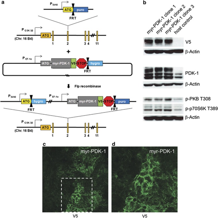
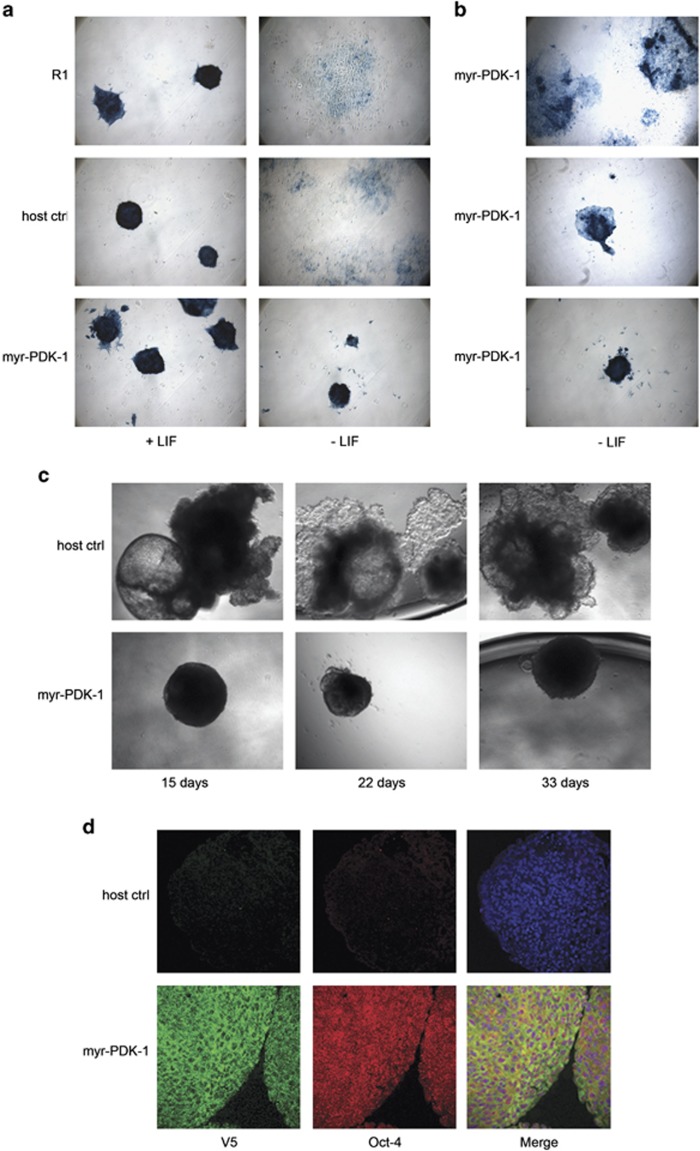
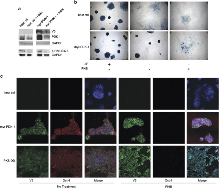
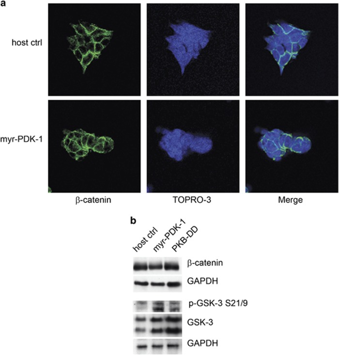
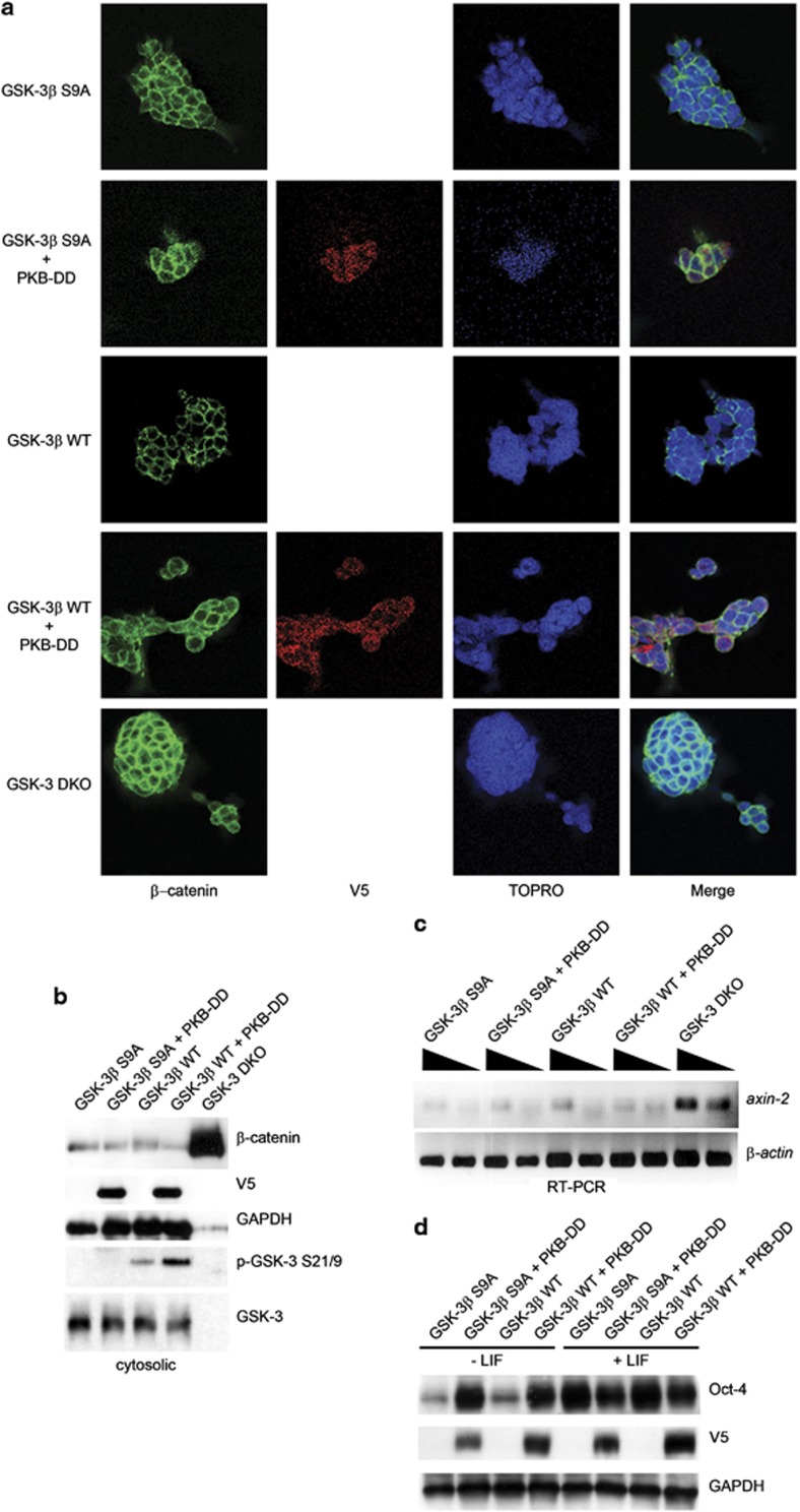
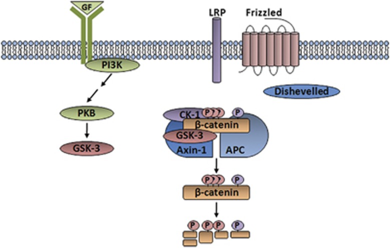
The phosphatidylinositol 3' kinase (PI3K) pathway is involved in many cellular processes including cell proliferation, survival and glucose transport, and is implicated in various disease states, such as cancer and diabetes. Although there have been numerous studies dissecting the role of PI3K signaling in different cell types and disease models, the mechanism by which PI3K signaling regulates embryonic stem (ES) cell fate remains unclear. It is believed that in addition to proliferation and tumorigenesis, PI3K activity may also be important for ES cell self-renewal. Paling et al. reported that the inhibition of PI3K led to a reduction in the ability of leukemia inhibitory factor to maintain self-renewal, causing cells to differentiate. Studies in our lab have revealed that ES cells completely lacking glycogen synthase kinase-3 (GSK-3) remain undifferentiated compared with wild-type ES cells. GSK-3 is negatively regulated by PI3K, suggesting that PI3K may have a vital role in maintaining pluripotency in ES cells through GSK-3. By using a modified Flp recombinase system, we expressed activated alleles of 3-phosphoinositide-dependent protein kinase-1 and protein kinase B to create stable, isogenic ES cell lines to further study the role of the PI3K signaling pathway in stem cell fate determination. In vitro characterization of the transgenic cell lines revealed a strong tendency toward the maintenance of pluripotency, and this phenotype was found to be independent of canonical Wnt signal transduction. In summary, PI3K signaling is sufficient to maintain the self-renewal and survival of stem cells. As this pathway is frequently mutationally activated in cancers, its effect on suppressing differentiation may contribute to its oncogenicity.


Figures

Figure 1
Figure 1
Modified Flp-In system. (a) A targeting vector is introduced to knock out a single allele of GSK-3β and create a host cell line with a FRT-puro cassette. Co-transfection of Flp recombinase and a gene of interest previously cloned into a vector containing a V5 epitope and FRT-hygro cassette results in homologous recombination at the FRT sites. Positive colonies are resistant to hygromycin and sensitive to puromycin. (b) Western blot analysis of transgenic myr-PDK-1 cell lines. (c) Membrane localization of myr-PDK-1 by immunofluorescent staining of V5; confocal, × 40 magnification.
Figure 2
Figure 2
Analysis of pluripotent markers on myr-PDK-1 cells. (a, b) Alkaline phosphatase activity. Cells were grown in monolayer and stained for alkaline phosphatase activity on day 5; phase contrast, × 10 magnification. (c) Gross morphology of embryoid bodies; phase contrast, × 5 magnification. (d) Oct-4 immunofluorescent expression of myr-PDK-1 transgenic embryoid bodies at day 10; confocal, × 40 magnification. Ctrl, control.
Figure 3
Figure 3
Treatment of myr-PDK-1 ES cells with PKB inhibitor. (a) Western blot analysis of whole-cell lysates after growing cells long term in inhibitor. (b) Cells grown in monolayer stained for alkaline phosphatase activity on day 5; phase contrast, × 10 magnification. (c) Immunofluorescent staining of cells grown in monolayer after 8 h treatment with PKBi; confocal, × 40 magnification. Ctrl, control; GAPDH, glyceraldehyde 3-phosphate dehydrogenase.
Figure 4
Figure 4
Treatment of myr-PDK-1ES cells with rapamycin (rapa). (a) Immunofluorescent staining of cells grown in monolayer after 8 h stimulation with rapa; confocal, × 40 magnification. (b) Western blot analysis of whole-cell lysates. Ctrl, control; GAPDH, glyceraldehyde 3-phosphate dehydrogenase.
Figure 5
Figure 5
Analysis of β-catenin expression in myr-PDK-1 and PKB-DD cells. (a) Immuno-fluorescent staining of myr-PDK-1 cells with β-catenin. (b) Western blot analysis of myr-PDK-1 and PKB-DD cytosolic cell lysates for β-catenin protein expression and whole-cell lysates for phospho-GSK-3 and pan-GSK-3 levels. Ctrl, control; GAPDH, glyceraldehyde 3-phosphate dehydrogenase.
Figure 6
Figure 6
Examination of cross talk between PI3K and Wnt signaling via GSK-3. (a) Analysis of PKB-mediated regulation of β-catenin expression via GSK-3 by immunofluorescent staining following transient transfection with PKB-DD-V5. (b) Western blot analysis of cytosolic cell lysates after transient transfection with PKB-DD-V5. (c) Semi-quantitative reverse transcription–PCR for axin-2 (1 × and 10 × template dilutions) after transient transfection with PKB-DD. (d) Western blot analysis of whole-cell lysates of GSK-3β S9A and GSK-3β WT cells transiently transfected with myr-PDK-1-V5. GAPDH, glyceraldehyde 3-phosphate dehydrogenase.
Figure 7
Figure 7
Immunohistology of 3-week-old teratomas. (a) Teratomas are stained with hematoxylin and eosin stain. Teratomas appear purple and the surrounding muscle fibers pink. Host control (Ctrl) teratomas are small and remain encapsulated, whereas myr-PDK1 teratomas grow into the surrounding muscle tissue. Whole slide scanning, scale bar=500 μm. (b) Teratomas were stained for V5 (brownish red) to illustrate areas of transgene expression. myr-PDK1 and PKB-DD expression is patchy throughout the teratoma. Scale bar=100 μm. (c) An equal number of cells were plated and allowed to grow in standard tissue culture conditions. Cells were counted on days 3 and 5. (d) Immunohistological staining of Ki67 and cleaved Caspase-3 (brown) in teratomas. Scale bar=100 μm.
Figure 8
Figure 8
Separate PI3K and Wnt signaling pathways. GSK-3 is a key regulator in both the PI3K and Wnt signal transduction pathways. In the context of the myr-PDK-1 and PKB-DD transgenic ES cell lines, the PI3K signaling pathway does not cross talk with the canonical Wnt signaling pathway through GSK-3. APC, adenomatous polyposis coli; LRP, lipoprotein-related protein; GF, growth factor.

References

    1. Hawkins PT, Anderson KE, Davidson K, Stephens LR. Signalling through class I PI3Ks in mammalian cells. Biochem Soc Trans. 2006;34 (Pt 5:647–662. - PubMed
    1. Engelman JA, Luo J, Cantley LC. The evolution of phosphatidylinositol 3-kinase as regulators of growth and metabolism. Nat Rev Gen. 2006;7:606–619. - PubMed
    1. Bader AG, Kang S, Zhao L, Vogt PK. Oncogenic PI3K deregulates transcription and translation. Nat Rev Cancer. 2005;5:921–929. - PubMed
    1. Scheid MP, Woodgett JR. PKB/AKT: functional insights from genetic models. Nat Rev Mol Cell Biol. 2001;2:760–768. - PubMed
    1. Zhao JJ, Wang L, Shin E, Loda MF, Roberts TM. The oncogenic properties of mutant p110alpha and p110beta phosphatidylinositol 3-kinases in human mammary epithelial cells. Proc Natl Acad Sci USA. 2005;102:18443–18448. - PMC - PubMed

Publication types

MeSH terms

Substances