. 2013 Apr;15(4):430-9.
doi: 10.1038/ncb2695. Epub 2013 Mar 3.

Ubiquitylation-dependent localization of PLK1 in mitosis

Affiliations

Ubiquitylation-dependent localization of PLK1 in mitosis

Jochen Beck et al. Nat Cell Biol. 2013 Apr.

Abstract

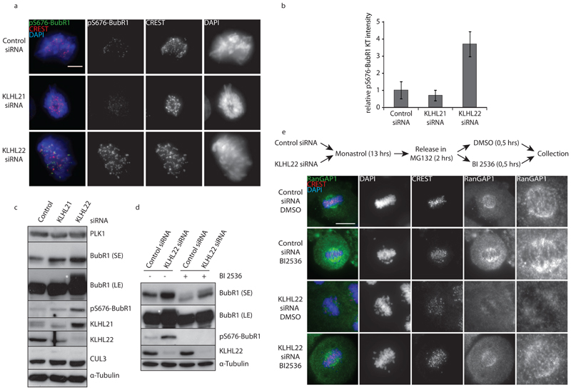
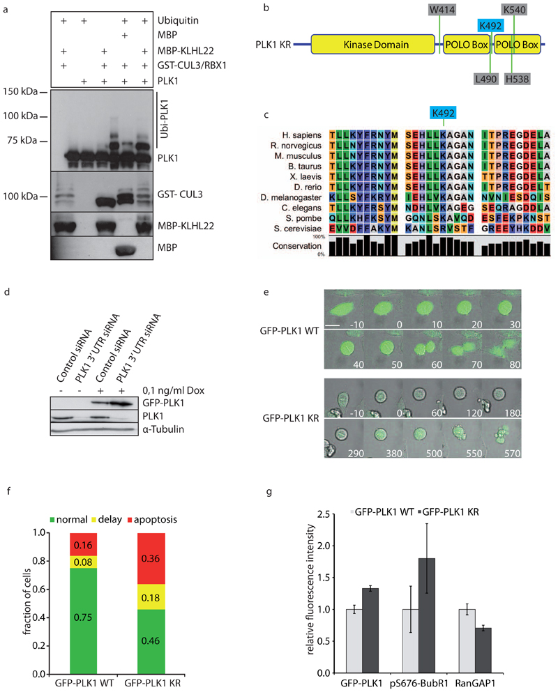
Polo-like kinase 1 (PLK1) critically regulates mitosis through its dynamic localization to kinetochores, centrosomes and the midzone. The polo-box domain (PBD) and activity of PLK1 mediate its recruitment to mitotic structures, but the mechanisms regulating PLK1 dynamics remain poorly understood. Here, we identify PLK1 as a target of the cullin 3 (CUL3)-based E3 ubiquitin ligase, containing the BTB adaptor KLHL22, which regulates chromosome alignment and PLK1 kinetochore localization but not PLK1 stability. In the absence of KLHL22, PLK1 accumulates on kinetochores, resulting in activation of the spindle assembly checkpoint (SAC). CUL3-KLHL22 ubiquitylates Lys 492, located within the PBD, leading to PLK1 dissociation from kinetochore phosphoreceptors. Expression of a non-ubiquitylatable PLK1-K492R mutant phenocopies inactivation of CUL3-KLHL22. KLHL22 associates with the mitotic spindle and its interaction with PLK1 increases on chromosome bi-orientation. Our data suggest that CUL3-KLHL22-mediated ubiquitylation signals degradation-independent removal of PLK1 from kinetochores and SAC satisfaction, which are required for faithful mitosis.


Conflict of interest statement

Competing financial interests

The authors declare no competing financial interests.

Figures

Figure 1
Figure 1. The BTB adaptor protein KLHL22 regulates SAC-dependent chromosome alignment during mitosis.
a, HeLa cells expressing H2B-mRFP were treated with the indicated siRNAs for 48 h and analyzed by live-cell microscopy. Stars indicate misaligned and lagging chromosomes. Bar is 10μm. b, The percentage of cells that complete mitosis normally or with a delay from prophase to anaphase (>50 min) or that undergo apoptosis during prometaphase was quantified (n = 50). c, The percentage of cells that undergo normal mitosis, become multinucleated or die during mitosis was quantified (n=50). d, HeLa cells were treated with the indicated siRNAs for 48 h and analyzed by immunofluorescence microscopy for MAD2 and kinetochores. The percentage of prometaphase and metaphase cells that contain one or more MAD2 positive kinetochores was quantified (n = 30). Example images are shown in Suppl. Fig. 1c. e, HeLa cells expressing H2B-mRFP were treated with the indicated mixtures of siRNAs for 48 h and analyzed by live-cell microscopy. The percentage of cells that die in prometaphase was quantified (n = 50). b-d, Bars represent the mean of three independent experiments. Error bars indicate +/-s.d.
Figure 2
Figure 2. The CUL3-KLHL22 E3-ligase interacts directly with PLK1 and regulates its kinetochore localization during mitosis.
a, HeLa cells inducibly expressing FLAG-KLHL22 were treated with doxycycline (Dox), synchronized by double thymidine block release and harvested in mitosis by mitotic shake-off. Cell extracts were immunoprecipitated with anti-FLAG antibodies and analyzed by Western blot. The asterisk indicates the band corresponding to PLK2. b, HeLa cells were synchronized as in (a) and extracts were immunoprecipitated with control (IgG IP) or anti-PLK1 (PLK1 IP) antibodies and analyzed by Western blot. c, Recombinant GST or GST fused to full length PLK1 (GST-PLK1) were incubated with recombinant MBP, MBP fused to full length KLHL21 (MBP-KLHL21) or KLHL22 (MBP-KLHL22), and immunoprecipitated using glutathione-sepharose beads. Immunoprecipitates (GST-IP) were washed in low and high salt conditions and analyzed by Coomassie blue staining and Western blot. d, Recombinant GST, GST fused to the Kelch repeats (GST-6K) of KLHL21, KLHL22 or KLHL9 were incubated with recombinant PLK1, immunoprecipitated using glutathionesepharose beads and analyzed by Western blot. e, HeLa cells were treated with the indicated siRNAs for 48 h, arrested in mitosis with Nocodazole for 14 h and analyzed by immunofluorescence microscopy. Bar is 5 μm. f, Quantification of relative intensity ratios of PLK1 : CREST staining on individual kinetochores (from 10 cells) from unsynchronized (as in Suppl. Fig. 3c, light grey) and Nocodazole synchronized cells(as in (e), dark grey). The ratios of control siRNA treated cells were set to 1. g, Quantification of intensity ratios of different kinetochore markers normalized to the corresponding CREST signals in control (light grey) and KLHL22 depleted (dark grey) cells. h, The relative kinetochore intensities of PLK1 were quantified as in (f) from cells treated with indicated siRNAs and Nocodazole (NOC), Taxol (TAX), Monastrol (MON), MG132 or DMSO. f, g, and h, Bars represent the mean of three independent experiments. Error bars indicate +/-s.d.
Figure 3
Figure 3. KLHL22 regulates PLK1-mediated phosphorylation of BubR1 and stable KTMT attachments.
a, HeLa cells were treated with the indicated siRNAs for 48 h, arrested in mitosis with Nocodazole for 14 h and analyzed by immunofluorescence microscopy. Bar 5 is μm. b, Quantification of the relative intensity ratios of pS676 BubR1 : CREST on individual kinetochores (n = 10) from (a). Bars represent the mean of three independent experiments. Error bars indicate +/-s.d. c, HeLa cells were treated as in (a), harvested by mitotic shake-off and analyzed by Western blot. d, HeLa cells were treated as in (c), incubated with BI 2536 as indicated and analyzed by Western blot. e, HeLa cells were treated with the indicated siRNAs for 48 h and analyzed by immunofluorescence microscopy. Insets are 3x zooms of the framed region. Bar is 10 μm.
Figure 4
Figure 4. CUL3-KLHL22-mediated ubiquitination of PLK1 within the PBD domain regulates faithful chromosome alignment during mitosis.
a, Recombinant PLK1 was added to in vitro ubiquitination reactions containing GSTCUL3/RBX1 complexes purified from Sf9 cells. Reactions were incubated for 1 h with MBP or MBP-KLHL22 purified from E. coli and analyzed by Western blot. Note that PLK1 was mainly monoubiquitinated (asterisk) under these conditions, but multiple sites were used with lower efficiency. b, Schematic representation of PLK1. The ubiquitin acceptor lysine K492 is indicated in blue, while residues required for PBD phosphosite targeting are indicated in grey.c, Alignment of PLK1 regions containing the identified ubiquitin acceptor site K492 from different species. d, HeLa cells inducibly expressing GFP-PLK1 WT were treated with doxycycline (+/- Dox) and transfected with control or PLK1 3’UTR siRNA. Cell extracts were analyzed by Western blot. e, HeLa cell lines inducibly expressing GFP-PLK1 wild type (WT) or the KR mutant were transfected with PLK1 3’UTR siRNA and analyzed by live-cell microscopy 24-48 h after transfection. Minutes before and after mitotic entry are indicated. Bar is 10μm. f, Quantification of cells from (e) that complete mitosis (green and yellow) versus cells that undergo apoptosis during prometaphase (red) (total numbers of cells analyzed: nWT=312, nKR=235). For the cells that complete mitosis, the duration from prophase to anaphase was quantified and the fraction of cells displaying a normal (green) or delayed (yellow) anaphase onset was calculated. Delay is defined as ‘duration longer than WT average + 1x standard deviation’. Duration values were quantified from nWT=80, nKR=78 cells. g, Cells expressing GFP-PLK1WT (light grey) or GFP-PLK1KR (dark grey) were transfected with PLK1 3’UTR siRNA, fixed 36 h after transfection and analyzed by immunofluorescence microscopy. GFP-PLK1 was visualized by GFP microscopy, pS676 BubR1, RanGAP1 and CREST were stained using the corresponding antibodies. To quantify kinetochore-associated GFP-PLK1, GFP at individual kinetochores (n=322 kinetochores in total from 3 experiments) was normalized both to cytoplasmic GFP and to the corresponding CREST signal at kinetochores using the following equation: GFP(kinetochore) / GFP(cytoplasm) / CREST(kinetochore). pS676 BubR1 signals of individual kinetochores (n=36 kinetochores from 1 experiment) were normalized both to the corresponding CREST and GFP-PLK1 signal intensities using the following equation: (pS676 BubR1/CREST)/(GFP/CREST), while RanGAP1 intensities on individual kinetochores (n=322 kinetochores in total from 3 experiments) were normalized to CREST staining. Error bars indicate +/-s.d.
Figure 5
Figure 5. CUL3-KLHL22-mediated ubiquitination of PLK1 regulates PBD-mediated phospho-interactions at kinetochores and stable KT-MT attachments.
a, HeLa cells inducibly expressing HIS-PLK1 WT or the KR mutant were transfected with PLK1 3’UTR siRNA and analyzed by immunofluorescence microscopy. 5x zooms of the boxed regions are shown on the right side. Note: expression of PLK1KR mutant leads to increase in kinetochore association of PLK1 and decrease in RanGAP1 at kinetochores but not at the nuclear envelope (arrows) in interphase cells. Bar is 5 μm. b, Cells expressing HISPLK1WT (light grey) or HIS-PLK1KR (dark grey) were treated as in (Fig 4 g) and kinetochoreassociated HIS-PLK1 (n=150 kinetochores in total from 3 experiments) and RanGAP1 (n=170 kinetochores in total from 3 experiments) were quantified as in (Fig 4 g). Error bars indicate +/-s.d. c, Model of PLK1 crystal structure highlighting the relevant residues of this study. The ubiquitin acceptor lysine K492 is shown in blue, the residues required for phosphopeptide targeting are shown in grey. d, Cells expressing GFP alone, GFP-PLK1WT or GFP-PLK1KR were synchronized in mitosis, immunoprecipitated using GFP-Trap beads and analyzed by Western blot. e, Cells expressing GFP alone, GFP-PLK1WT or GFP-PLK1KR were transfected with either HA alone or HA-KLHL22 and immunoprecipitated using GFPTrap beads and analyzed by Western blot.
Figure 6
Figure 6. KLHL22 accumulates at the mitotic spindle and its association with PLK1 peaks with chromosome biorentation.
a, Immunofluorescence microscopy was performed using a Doxycycline-inducible GFP-KLHL22 cell line, which was synchronized in mitosis by RO3306 block and release, fixed and stained with CREST and α-tubulin using specific antibodies. GFP-KLHL22 is visualized only by its GFP fluorescence. Images are maximum intensity projections through Z-stacks spanning a total depth of 12 μm in 0.5μm increments. Bar is 10 μm. b, Workflow for synchronizing cells in metaphase and addition of DMSO, the microtubule-stabilizing drug Taxol (TAX) or the microtubule-depolymerizing agent Nocodazole (NOC). Note that Taxol allows MT attachment without tension being exerted on kinetochores, while kinetochores in Nocodacole-treated cells are not attached to MT’s. c, HeLa cells expressing HA-KLHL22 and GFP-PLK1 were treated as outlined in (b), harvested and GFP-PLK1 was immunoprecipitated and analyzed by Western blot. d, HeLa cells expressing HA-KLHL22 were treated as outlined in (b), harvested and endogenous PLK1 was immunoprecipitated and analyzed by Western blot. e, Model for ubiquitination-dependent regulation of PLK1 in mitosis by CUL3/KLHL22. PLK1 (green) localizes to kinetochores (dark blue) and phosphorylates (P) the key SAC component, BubR1 kinase (beige) to control chromosome alignment during prometaphase and metaphase stages. Following the establishment of stable attachments of kinetochores by microtubules (Tension) (black bars), CUL3 (red) together with the substrate specific adaptor protein KLHL22 (light blue) binds and ubiquitinates PLK1 (Ub, yellow) within the PBD domain leading to its dissociation from phosphoreceptor proteins and thereby efficient removal from kinetochores, allowing for silencing of SAC and chromosome segregation. This process may occur dynamically during prometaphase stages to allow for a precise correction mechanism sensing stable MT-KT interactions and microtubule-exerted tension and satisfaction of the SAC.

Comment in

Similar articles

Cited by

References

    1. Williams BR, Amon A. Aneuploidy: cancer's fatal flaw? Cancer Res. 2009;69(13):5289–5291. - PMC - PubMed
    1. Lengauer C, Kinzler KW, Vogelstein B. Genetic instabilities in human cancers. Nature. 1998;396(6712):643–649. - PubMed
    1. Strebhardt K. Multifaceted polo-like kinases: drug targets and antitargets for cancer therapy. Nat Rev Drug Discov. 2010;9(8):643–660. - PubMed
    1. Petronczki M, Lenart P, Peters JM. Polo on the Rise-from Mitotic Entry to Cytokinesis with Plk1. Dev Cell. 2008;14(5):646–659. - PubMed
    1. Sumara I, et al. Roles of polo-like kinase 1 in the assembly of functional mitotic spindles. Curr Biol. 2004;14(19):1712–1722. - PubMed

Publication types

MeSH terms