. 2013 Apr 1;190(7):3639-47.
doi: 10.4049/jimmunol.1203488. Epub 2013 Mar 1.

NKG2D blockade inhibits poly(I:C)-triggered fetal loss in wild type but not in IL-10-/- mice

Affiliations

NKG2D blockade inhibits poly(I:C)-triggered fetal loss in wild type but not in IL-10-/- mice

Jessica E Thaxton et al. J Immunol. .

Abstract

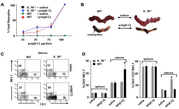
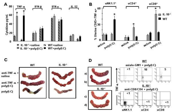
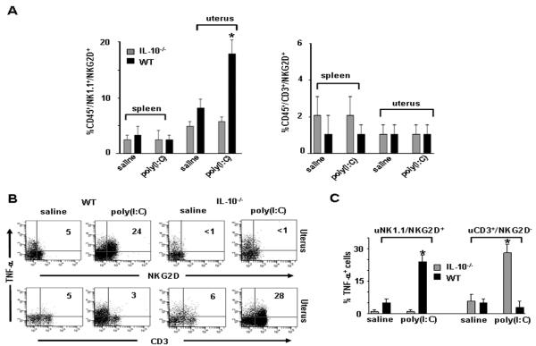
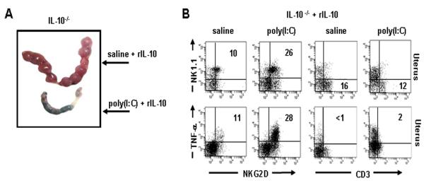
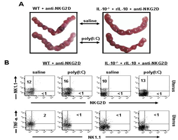
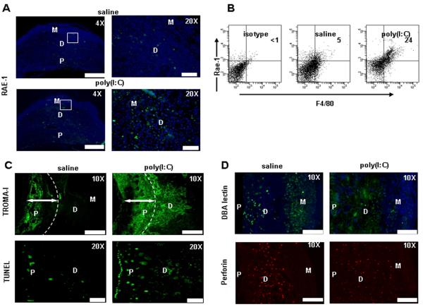
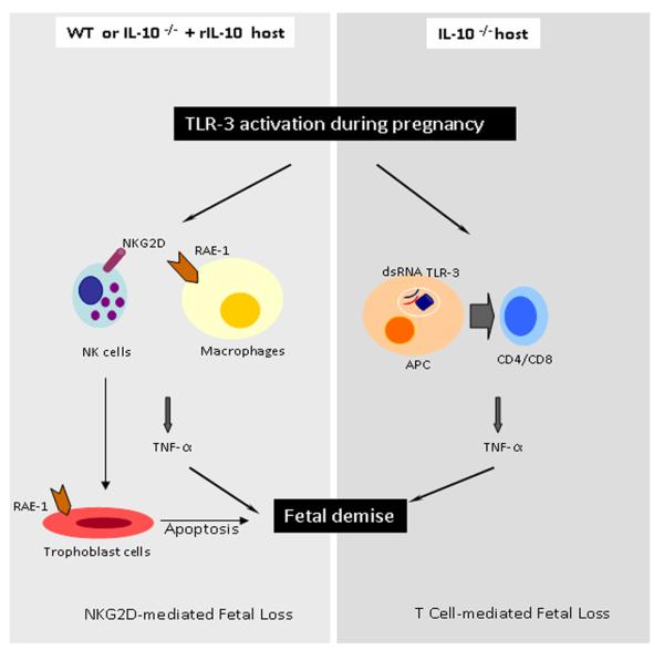
Infection and inflammation can disturb immune tolerance at the maternal-fetal interface, resulting in adverse pregnancy outcomes. However, the underlying mechanisms for detrimental immune responses remain ill defined. In this study, we provide evidence for immune programming of fetal loss in response to polyinosinic:polycytidylic acid (polyI:C), a viral mimic and an inducer of inflammatory milieu. IL-10 and uterine NK (uNK) cells expressing the activating receptor NKG2D play a critical role in poly(I:C)-induced fetal demise. In wild type (WT) mice, poly(I:C) treatment induced expansion of NKG2D(+) uNK cells and expression of Rae-1 (an NKG2D ligand) on uterine macrophages and led to fetal resorption. In IL-10(-/-) mice, NKG2D(-) T cells instead became the source of fetal resorption during the same gestation period. Interestingly, both uterine NK and T cells produced TNF-α as the key cytotoxic factor contributing to fetal loss. Treatment of WT mice with poly(I:C) resulted in excessive trophoblast migration into the decidua and increased TUNEL-positive signal. IL-10(-/-) mice supplemented with recombinant IL-10 induced fetal loss through NKG2D(+) uNK cells, similar to the response in WT mice. Blockade of NKG2D in poly(I:C)-treated WT mice led to normal pregnancy outcome. Thus, we demonstrate that pregnancy-disrupting inflammatory events mimicked by poly(I:C) are regulated by IL-10 and depend on the effector function of uterine NKG2D(+) NK cells in WT mice and NKG2D(-) T cells in IL-10 null mice.


Figures

Figure 1
Figure 1. Fetal resorption and amplification of uterine NK and T cells in WT and IL-10−/− mice in response to poly(I:C) treatment
(A) poly(I:C) injected i.p. on gd6 was evaluated in a dose dependent manner to induce fetal resorption as assessed by inspection of uterine placental units on gd10. A dose of 100 μg/mouse induced 100% fetal resorption in both IL-10−/− and WT mice. A subset of these mice was allowed to deliver and no pups were born. Data are plotted as mean ± S.E.M (n=6/treatment). (B) Representative gd10 WT and IL-10−/− uterine horns from mice treated with saline or 100 μg/mouse poly(I:C) are depicted. (C) Assessment of splenic and uterine immune cells from WT or IL-10−/− mice treated on gd6 with saline or poly(I:C) (100 μg/mouse) and harvested on gd10. Cellular populations were first gated on CD45+ cells then analyzed for NK1.1 versus CD3. Data from spleen and uterus are representative of 8 mice per condition and numbers are averages of these data. (D) Graphs indicate statistical significance (*, p<0.05) of saline versus poly(I:C) treated cellular populations as indicated.
Figure 2
Figure 2. Measurement of cytokines and effect of in vivo TNF-α neutralization on fetal resoprtion in WT Mice and IL-10−/− mice
(A) ELISA was performed using gd10 serum samples from IL-10−/− or WT mice treated on gd6 with saline or poly(I:C) (100 μg/mouse) and the analysis included TNF-α, IFN-β, IFN-γ, and IL-12. Bars represent mean ± S.E.M. (n=8 mice/group) and (*; p<0.05) indicates significance between saline and poly(I:C) treatments. (B) FACS analysis of TNF-α producing uterine NK1.1, CD4+ and CD8+ cells. Uterine mononuclear cells were isolated from gd10 uteroplacental tissue and gated for CD45 positive staining and further analyzed for NK1.1, CD4+CD8+ subpopulations and TNF-α expression. Graphic representation is provided from three independent experiments for intracellular staining of TNF-α produced by the indicated cellular populations in WT or IL-10−/− mice treated with saline or poly(I:C) on gd6. Data are plotted as mean ± S.E.M. (n=9 mice/condition) and (*) indicates statistical significance (p<0.05) between saline and poly(I:C) treated samples. (C) Representative gd10 uterine horns from WT (left) or IL-10−/− (right) mice treated with (from top to bottom of panels) saline + anti-TNF-α antibody, poly(I:C) + anti-TNF- α antibody, or poly(I:C) + isotype antibody (n=4 mice/ condition). (D) Rescue of pregnancy in response to NK cell depletion by asialo-GM1 antibody and T cell depletion by anti-CD4 and anti-CD8 antibodies. a) gd10 uterine horns from WT mice treated with asialo-GM1 antibody + saline, b) asialo-GM1 antibody + poly(I:C), c) gd10 uterine horns from IL-10−/− mice treated with anti-CD4 antibody + anti-CD8 antibody + saline, and d) anti-CD4 antibody + anti-CD8 antibody + poly(I:C). Corresponding dot plots represent data from n=4 independent experiments where TNF-α production was assessed from the cell populations indicated under depletion conditions featured in (b-d).
Figure 3
Figure 3. Induction of NKG2D in NK1.1+/TNF- α+ uNK cells in WT mice in response to poly(I:C)
(A) Representative plots of splenic or uterine CD45+NK1.1+NKG2D+ and CD45+CD3+NKG2D+ cells as analyzed by FACS from gd10 WT or IL-10−/− mice treated on gd6 with saline or poly(I:C). Graphs are average of cell populations from 8 mice per condition, (*; p<0.05) indicates significance between saline and poly(I:C) treatment conditions. (B) Representative FACS analysis of intracellular TNF-α in uterine CD45+ cells gated on the NKG2D+ and CD3+ subpopulations from gd10 WT or IL-10−/− mice treated on gd6 with saline or poly(I:C). (C) Summary graphs of TNF-α+ cells gated NK1.1+/NKG2D+ and CD3+/NKG3D isolated from uterine tissue (n=8 mice/ condition). *; p<0.05 shows significance between saline and poly(I:C) treatment conditions.
Figure 4
Figure 4. Recombinant IL-10 (rIL-10) induces NKG2D on uterine NK1.1 cells in IL-10−/− mice
(A) Representative gd10 uterine horns from IL-10−/− mice treated with saline or poly(I:C) + rIL- 10 (n=4 mice/condition). rIL-10 supplementation does not protect against fetal resorption. (B) Uterine populations were analyzed by FACS from IL-10−/− mice supplemented with rIL-10 and treated with saline or poly(I:C). Data represent mean ± S.E.M. (n= 4 mice/condition). IL-10−/− mice supplemented with rIL-10 show NKG2D expression on NK1.1 cells which now produce TNF-α.
Figure 5
Figure 5. Depletion of NKG2D+ NK cells rescues pregnancy and leads to loss of TNF-α production in WT and rIL-10 supplemented IL-10−/− mice in response to poly(I:C)
(A) Representative uterine horns from gd10 WT or rIL-10-supplemented IL-10−/− mice treated with anti-NKG2D antibody + saline or anti-NKG2D antibody + poly(I:C). Data represent mean ± S.E.M. (n= 4 mice/condition). NKG2D depletion abrogated fetal resorption as demonstrated by normal fetal units. (B) Assessment of NKG2D depletion and loss of TNF-α production was performed by gating on CD45+NK1.1+NKG2D+ uterine cells from saline or poly(I:C)-treated gd10 WT or rIL-10 + IL-10−/− mice injected i.p. with anti-NKG2D antibody. Dot plots represent data from 4 or more animals per condition.
Figure 6
Figure 6. Exposure of pregnant WT mice to poly(I:C) results in induced expression of Rae- 1 on macrophages and enhanced trophobalst invasion and cell death
(A) Representative images from uteroplacental units from gd10 saline or poly(I:C) treated WT mice was assessed by immunohistochemistry for RAE-1 expression. Weak staining for Rae-1 was detected in the decidua and placental regions (4× magnification; Scale Bar: 100 μm and 20× magnification; Scale Bar: 20 μm). (B) FACS analysis of Rae-1 on uterine immune cells. CD45+ and CD45cytokeratin7+ cells were analyzed. Cells are gated from CD45+ populations obtained from uterine populations. Data represent three independent experiments. (C) Upper panel: immunohistochemical staining with TROMA-I antibody shows unique staining with increased invasion of trophoblast cells into the decidua in response to poly(I:C), but not saline (10× magnification; Scale Bar: 60 μm). Lower panel: TUNEL staining (20× magnification; Scale Bar: 20 μm) demonstrates significantly increased cell death the trophoblastic regions, including invading trophoblasts. Images are representative of a total of 3mice per condition. (D) DBA+ and PRF+ NK cell identification by immunofuorescence staining from uteroplacental unit from gd10. In response to poly(I:C) treatment, DBA+ intensity is diminished but these cells maintain their PRF+ phenotype. However, their distribution appears to be diffused through the placenta (P) region. Images are representative of results obtained from placental samples of 3 mice per condition (10xmagnification; Scale Bar: 60 μm).
Figure 7
Figure 7. Schematic representation of the poly(I:C)-induced events in WT and IL-10−/− mice
The model recapitulates the pathways leading to uterine NK or T cell-mediated cytotoxicity and fetal resorption in WT and IL-10−/− mice, respectively. Rae-1 positive macrophages or trophoblasts can interact with NKG2D+ NK cells and induce TNF-α production and trophoblast cell death in response to poly(I:C). On the other hand, uterine T cells can be activated by poly(I:C) in the absence of IL-10 to produce TNF-α and to cause fetal demise.

Similar articles

Cited by

References

    1. Goldenberg RL, Hauth JC, Andrews WW. Intrauterine infection and preterm delivery. N. Engl. J. Med. 2000;342:1500–1507. - PubMed
    1. Haun L, Kwan N, Hollier LM. Viral infections in pregnancy. Minerva. Ginecol. 2007;59:159–174. - PubMed
    1. Romero R, Espinoza J, Chaiworapongsa T, Kalache K. Infection and prematurity and the role of preventive strategies. Semin. Neonatol. 2002;7:259–274. - PubMed
    1. Thaxton JE, Nevers TA, Sharma S. TLR-mediated preterm birth in response to pathogenic agents. Infect. Dis. Obstet. Gynecol. 2010;2010 pii.378472. - PMC - PubMed
    1. Moffet A, Loke C. Immunology of placentation in eutharian mammals. Nat. Rev. Immunol. 2006;8:584–594. - PubMed

Publication types

MeSH terms

LinkOut - more resources