. 2013 Mar:Chapter 14:Unit14.23.
doi: 10.1002/0471141755.ph1423s60.

Patient-derived models of human breast cancer: protocols for in vitro and in vivo applications in tumor biology and translational medicine

Affiliations

Patient-derived models of human breast cancer: protocols for in vitro and in vivo applications in tumor biology and translational medicine

Yoko S DeRose et al. Curr Protoc Pharmacol. 2013 Mar.

Abstract

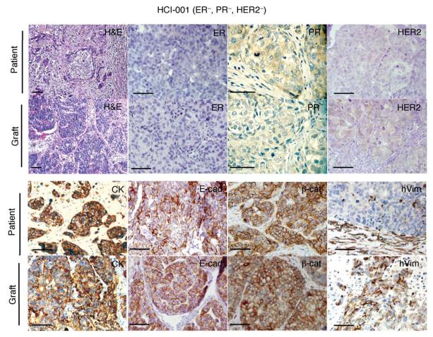
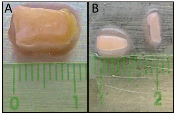
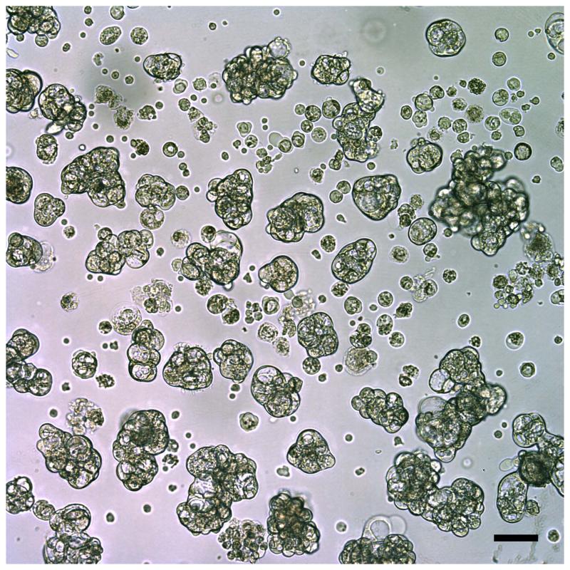
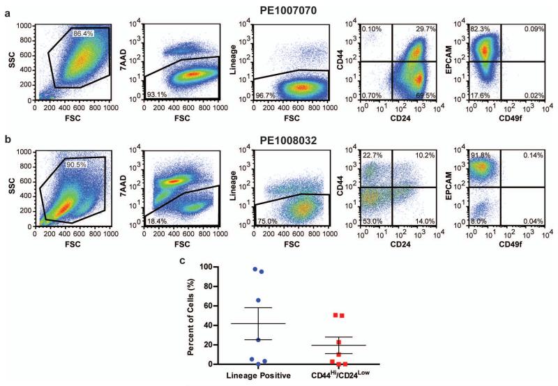
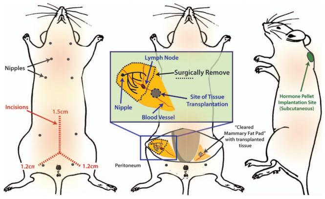
Research models that replicate the diverse genetic and molecular landscape of breast cancer are critical for developing the next-generation therapeutic entities that can target specific cancer subtypes. Patient-derived tumorgrafts, generated by transplanting primary human tumor samples into immune-compromised mice, are a valuable method to model the clinical diversity of breast cancer in mice, and are a potential resource in personalized medicine. Primary tumorgrafts also enable in vivo testing of therapeutics and make possible the use of patient cancer tissue for in vitro screens. Described in this unit are a variety of protocols including tissue collection, biospecimen tracking, tissue processing, transplantation, and three-dimensional culturing of xenografted tissue, which enable use of bona fide uncultured human tissue in designing and validating cancer therapies.


Figures

Figure 1
Figure 1
Tumorgrafts resembled the original tumors from which they were derived. A representative ERPRHER2 tumor graft (HCI—001) is shown in comparison to the original patient sample. The tumor ID and the original clinical diagnosis for ER, PR, and HER2 are shown at the top. Sections from the patient’s primary breast tumor (patient), and from representative tumor grafts from the same patient (graft). Stains shown are hematoxylin and eosin (H&E) as well as antibody stains for ER, PR, HER2, cytokeratin (CK), E—cadherin (E—cad), β—catenin (β—cat), and human specific vimentin (hVim). Positive antibody signals are brown in color, with hematoxylin (blue) counterstain. Some images are shown at higher magnification to visualize nuclear staining. All scale bars correspond to 100 μm. This figure was reprinted from DeRose et al., 2011 with permission from Nature Medicine.
Figure 2
Figure 2
Example of breast tumor tissue isolated from a patient. A. Surgical sample containing tumor tissue (white-pink color) and fat (yellow color). B. Fragments of the same tissue sample following removal of the fat and dissection into approximately 4 mm × 2 mm pieces, ready for implantation into mammary fat pads. The halo surrounding the tissue is due to the fluid used to keep the samples moist during processing.
Figure 3
Figure 3
Metastatic tumor cells collected from a pleural effusion and viewed using light microscopy. These breast tumor cells aggregated into organoid-like structures, which were easily separated from single cells such as leukocytes that were present in the original specimen. Scale bar is 100 μm.
Figure 4
Figure 4
(A) PE1007070 and (B) PE1008032, which are primary cells isolated from pleural effusions from two separate patients were analyzed by FACS for 7-AAD and Lineage markers in combination with either CD44/CD24 or EPCAM/CD49f. (C) Primary cells isolated from pleural effusions from seven patients were analyzed by FACS for Lineage markers and CD44/CD24.
Figure 5
Figure 5
Schematic showing location of mouse mammary fat pads, surgical incisions, and sites for tissue/cell transplants and the estrogen pellet implant. Mouse diagram was adapted from Hummel et al., 1966 with permission of The Jackson Laboratory.
Figure 6
Figure 6
Analysis of the viability of patient-derived breast cancer organoids in 3D culture following drug treatment. Primary tumor organoids were embedded in EHS matrix and grown in 3D culture. Organoids were treated with (A) DMSO or valproic acid for 4 days or (B) DMSO or taxol for 7 days and imaged using both DIC (bottom rows) and fluorescence microscopy (top rows). Live/dead discrimination was performed using calcein AM (green) to mark viable cells, and ethidium bromide homodimer-1 (red) to identify dead cells. Scale bars represents 20 μm. Panel A was adapted from Cohen, et al., 2011 with permission of Nature Publishing Company.

References

    1. Al-Hajj M, Wicha MS, Benito-Hernandez A, Morrison SJ, Clarke MF. Prospective identification of tumorigenic breast cancer cells. Proc. Natl. Acad. Sci. 2003;100:3983–3988. - PMC - PubMed
    1. Borowsky AD. Choosing a mouse model: Experimental biology in context—the utility and limitations of mouse models of breast cancer. Cold Spring Harbor Perspectives in Biology. 2011;3:a009670. - PMC - PubMed
    1. Burdall S, Hanby A, Lansdown M, Speirs V. Breast cancer cell lines: Friend or foe? Breast Cancer Res. 2003;5:89–95. - PMC - PubMed
    1. Caligiuri I, Rizzolio F, Boffo S, Giordano A, Toffoli G. Critical choices for modeling breast cancer in transgenic mouse models. J. Cell. Physiol. 2012;227:2988–2991. - PubMed
    1. Cespedes MV, Casanova I, Parreno M, Mangues R. Mouse models in oncogenesis and cancer therapy. Clin. Transl. Oncol. 2006;8:318–329. - PubMed

Publication types