Review
. 2013 May;113(1):1-11.
doi: 10.1007/s11060-013-1084-8. Epub 2013 Mar 2.

Biogenesis of extracellular vesicles (EV): exosomes, microvesicles, retrovirus-like vesicles, and apoptotic bodies

Affiliations
Review

Biogenesis of extracellular vesicles (EV): exosomes, microvesicles, retrovirus-like vesicles, and apoptotic bodies

Johnny C Akers et al. J Neurooncol. 2013 May.

Abstract

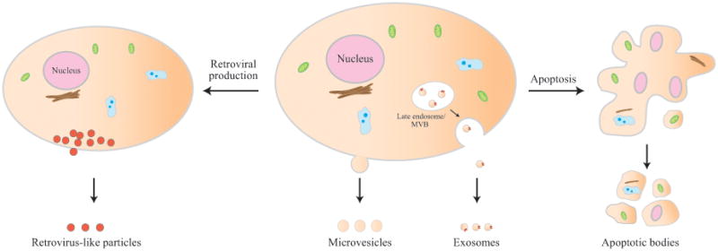
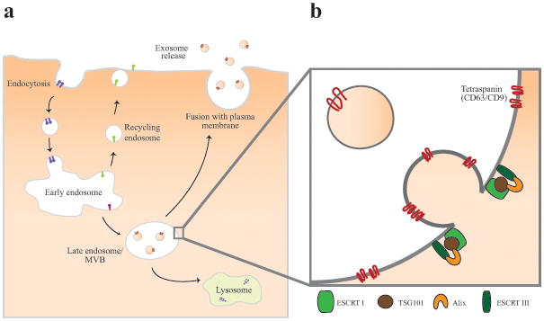
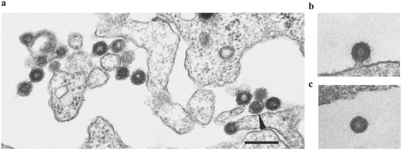
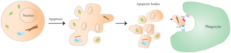
Recent studies suggest both normal and cancerous cells secrete vesicles into the extracellular space. These extracellular vesicles (EVs) contain materials that mirror the genetic and proteomic content of the secreting cell. The identification of cancer-specific material in EVs isolated from the biofluids (e.g., serum, cerebrospinal fluid, urine) of cancer patients suggests EVs as an attractive platform for biomarker development. It is important to recognize that the EVs derived from clinical samples are likely highly heterogeneous in make-up and arose from diverse sets of biologic processes. This article aims to review the biologic processes that give rise to various types of EVs, including exosomes, microvesicles, retrovirus like particles, and apoptotic bodies. Clinical pertinence of these EVs to neuro-oncology will also be discussed.


Figures

Fig 1
Fig 1
Biogenesis of the various types of extracellular vesicles: exosomes, microvesicles, retrovirus-like vesicles, and apoptotic bodies.
Fig 2
Fig 2
Electron micrograph of exosomes in maturing sheep reticulocytes. (a) Immunogold labeling with a monoclonal antibody against transferrin receptor. After eighteen hours of incubation, the gold label is found in the MVB but associated with the surface of the internal exosomes. The black arrow shows a sac beginning to fuse with the plasma membrane. (b) After 36 h of incubation, fusion is complete, and exosomes are released. Reprinted with permission from Blood Cells, Molecules, and Diseases [127].
Fig 3
Fig 3
Biogenesis and release of exosomes. (a) Exosomes are formed within the endosomal network. Early endosomes fuses with endocytic vesicles and incorporate their content into those destined for recycling, degradation, or exocytosis. Late endosomes, or multi-vesicular bodies (MVBs), develop from early endosomes, and are characterized by the presence of multiple small interluminal vesicles (ILVs). Exosomes are released from late endosomal compartment through the fusion of MVBs to the plasma membrane. (b) A key step in ILV formation is the reorganization of endosomal membrane protein such as CD9 and CD63 into tetraspanin enriched microdomains. Next, a series of Endosomal Sorting Complex Required for Transport, or ESCRTs are recruited to the site of budding. ESCRTI and II drive membrane budding and ESCRT III is required for completion of budding. ESCRTIII is recruited to the site of ESCRTI and II via Alix.
Fig 4
Fig 4
Microvesicle arises through outward budding and fission of plasma membrane and is the result of dynamic interplay between phospholipid redistribution and cytoskeletal protein contraction. Membrane budding/vesicle formation is induced by translocation of phosphatidylserine to the outer-membrane leaflet through the activity of aminophospholipid translocases. To enable microvesicle budding, ADP-ribosylation factor 6 (ARF6) initiates a signaling cascade that starts with the activation of phospholipase D (PLD), which recruits the extracellular signal-regulated kinase (ERK) to the plasma membrane. ERK phosphorylates and activates myosin light-chain kinase (MLCK). Phosphorylation and activation of the myosin light chain by MLCK triggers the release of microvesicles.
Fig 5
Fig 5
Electron micrograph of retrovirus-like particles budding from teratocarcinoma cell lines, GH (a) and Tera-1 (b, c). Scale bar = 250 nm. Reprinted with permission from Journal of General Virology [83].
Fig 6
Fig 6
Formation of apoptotic bodies during apoptosis. A cell dying by apoptosis progress through several stages, initiating with condensation of the nuclear chromatin, followed by membrane blebbing, progressing to disintegration of the cellular content into distinct membrane enclosed vesicles termed apoptotic bodies or apoptosomes. The clearance of apoptotic bodies by macrophages via phagocytosis is mediated by specific interactions between recognition receptors on the phagocytes and the specific changes in the composition of the apoptotic cell membrane. Theses changes include the oxidation of surface molecules, which create sites for binding of Thrombospondin (Tsp) or the complement protein C3b.

References

    1. Skog J, et al. Glioblastoma microvesicles transport RNA and proteins that promote tumour growth and provide diagnostic biomarkers. Nat Cell Biol. 2008;10(12):1470–6. - PMC - PubMed
    1. Raposo G, et al. B lymphocytes secrete antigen-presenting vesicles. J Exp Med. 1996;183(3):1161–72. - PMC - PubMed
    1. Blanchard N, et al. TCR activation of human T cells induces the production of exosomes bearing the TCR/CD3/zeta complex. J Immunol. 2002;168(7):3235–41. - PubMed
    1. Andre F, et al. Exosomes as Potent Cell-Free Peptide-Based Vaccine. I. Dendritic Cell-Derived Exosomes Transfer Functional MHC Class I/Peptide Complexes to Dendritic Cells. The Journal of Immunology. 2004;172(4):2126–2136. - PubMed
    1. Taylor DD, Akyol S, Gercel-Taylor C. Pregnancy-Associated Exosomes and Their Modulation of T Cell Signaling. The Journal of Immunology. 2006;176(3):1534–1542. - PubMed