. 2013 Sep;94(3):399-407.
doi: 10.1189/jlb.0512249. Epub 2013 Mar 1.

Pannexin1 hemichannels are critical for HIV infection of human primary CD4+ T lymphocytes

Affiliations

Pannexin1 hemichannels are critical for HIV infection of human primary CD4+ T lymphocytes

J A Orellana et al. J Leukoc Biol. 2013 Sep.

Abstract

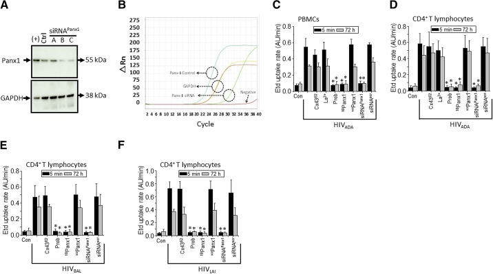
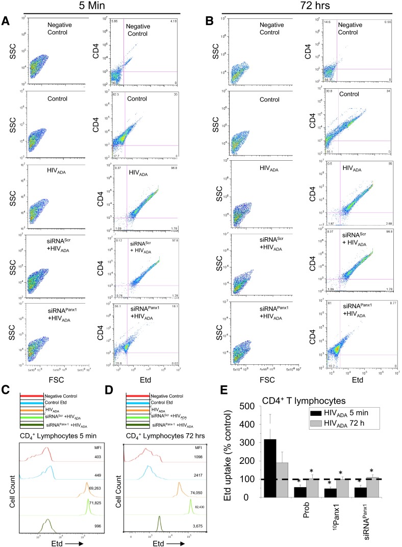
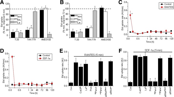
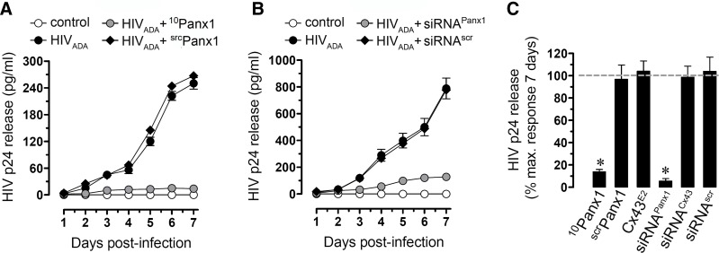
HIV is a major public health issue, and infection of CD4(+) T lymphocytes is one of its key features. Whereas several cellular proteins have been identified that facilitate viral infection and replication, the role of hemichannels in these processes has not been fully characterized. We now show that the HIV isolates, R5 and X4, induced a transient-early (5-30 min) and a later, persistent (48-120 h) opening of Panx1 hemichannels, which was dependent on the binding of HIV to CD4 and CCR5/CXCR4 receptors. Blocking Panx1 hemichannels by reducing their opening or protein expression inhibited HIV replication in CD4(+) T lymphocytes. Thus, our findings demonstrate that Panx1 hemichannels play an essential role in HIV infection.

Keywords: AIDS; channels; disease.


Figures

Figure 1.
Figure 1.. HIV increases Etd uptake in a biphasic manner in human primary PBMCs and CD4+ T lymphocytes.
(A–D) Time course of Etd uptake rate obtained from PBMCs or CD4+ T lymphocytes under control conditions (squares) or after infection with HIV (circles), 0–120 h. PBMCs infected with HIVADA (A) and CD4+ T lymphocytes infected with HIVADA (B), HIVBal (C), or HIVLAI (D). (E and F) Representative time-lapse measurements of Etd uptake in CD4+ T lymphocytes under control conditions (squares) or after 5 min (E) or 72 h (F) of exposure to HIVADA (circles). No differences were observed between R5 and X4 isolates. *P < 0.005; **P < 0.002 ; ***P = 0.0001 denote significance as compared with control conditions. Each value corresponds to the mean ± sd of the Etd intracellular intensity, present in at least 20 cells/time-point (n=4).
Figure 2.
Figure 2.. HIV increases the activity of Panx1 hemichannels in human primary CD4+ T lymphocytes.
(A) Total levels of Panx1 in CD4+ T lymphocytes under control conditions (Ctrl) or after transfection for 48 h with three different siRNAs (siRNA A, B, and C) to Panx1 hemichannels were analyzed by Western blot. HeLa cells transfected with Panx1 were used as a positive control of Panx1 protein expression [(+)]. Loading controls, analyzed by probing for GAPDH, are also shown. (B) Representative example of qRT-PCR curves of Panx1 mRNA expression in control (cyan line) and after transfection of siRNA to Panx1 (light green line). GAPDH was used as a control (yellow and red lines). The negative control without enzyme did not show any amplification (dark red line). (C and D) Etd uptake rate for PBMCs exposed to HIVADA (C); CD4+ T lymphocytes exposed to HIVADA (D), HIVBal (E), or HIVLAI (F) after 5 min (black bars) or 72 h (gray bars). (C–F) Control (Con), uninfected conditions are shown. Addition of Cx43 hemichannel blockers, Cx43E2 (1:500 dilution) and La3+ (200 μM), did not affect the Etd uptake rate induced by the virus. In contrast, Panx1 hemichannel blockers, Prob (500 μM) and 10Panx1 (200 μM), completely blocked Etd uptake rate induced by the virus. The negative control using a scrPanx1 peptide (200 μM) did not affect Etd uptake rate induced by the virus. Cells were transfected with siRNA C for Panx1 or with the appropriate siRNAscr (both 10 nM), and Etd uptake rate was analyzed. siRNA for Panx1, but not scramble control, reduced Etd uptake rate induced by the virus. Each value corresponds to the mean ± sd of the Etd intracellular intensity present in at least 20 cells/time-point (n=4; *P<0.005).
Figure 3.
Figure 3.. HIV exposure/infection of CD4+ T lymphocytes results in opening of Panx1 hemichannels as quantified by FACS.
(A and B) FACS analysis showing Etd uptake in CD4+ T lymphocytes after 5 min (A) or 72 h (B) of HIV exposure. (A and B, left) Side-scatter (SSC) versus forward-scatter (FSC) of the cells to demonstrate the purity and selection of the CD4+ population of lymphocytes; (right) CD4 staining versus Etd uptake for each condition. Unstained Etd conditions (Negative Control) showed no staining for Etd, whereas untreated cells (Control) showed minimal Etd uptake compared with HIVADA-treated cultures (HIVADA) for 5 min (A) or 72 h (B). Cells transfected with siRNA for Panx1 abolished the Etd-uptake increase induced by the virus at both time-points (A and B; siRNAPanx1+HIVADA). (C and D) Representative histograms of FACS staining using unstained cells (Negative Control, red lines), control conditions with Etd (blue lines), after exposure to HIVADA (orange lines), after siRNAscr + HIVADA (light green lines), or after siRNAPanx1 + HIVADA (dark green lines). MFI, Mean fluorescence intensity. (E) Quantification of data from five independent experiments using FACS analysis showed that blocking of Panx1 hemichannels with Prob, 10Panx1, or siRNAPanx1 reduced Etd uptake induced by HIVADA in CD4+ T lymphocytes. Each value corresponds to the mean ± sd of the Etd intracellular intensity, present in at least 20 cells/time-point (n=4 independent CD4+ T lymphocyte isolations; *P<0.005).
Figure 4.
Figure 4.. The HIV-induced Panx1 hemichannel opening occurs by a CD4 and a CCR5/CXCR4-binding/fusion-dependent mechanism.
A fusion inhibitor (T-20), CCR5 blocker (TAK-779), or CXCR4 blocker (AMD3100) on Etd uptake, induced by HIVADA (black bars), HIVBal (white bars), and HIVLAI (gray bars) infection at 5 min (A) and 72 h (B) of exposure. Data are presented as percent of maximal response of Etd uptake (% max. response). (C and D) Etd uptake rate obtained from CD4+ T lymphocytes under control conditions (squares) or after a single addition of the following chemokines (circles): RANTES/CCL5 (C) or SDF-1α/CXCL12 (D). (E and F) The effects on Etd uptake of the Cx43 hemichannel blockers, Cx43E2 (1:500 dilution) and La3+ ion (200 μM), or Panx1 hemichannel blockers, Prob (500 μM), 10Panx1 (200 μM), siRNA to Panx1, as well as negative control scrPanx1 (200 μM) or siRNAscr, are shown (E and F; represent 5 min after chemokine treatment). *P < 0.005. Each value corresponds to the mean ± sd of Etd intracellular intensity in at least 20 cells/time-point (n=4 for all experiments).
Figure 5.
Figure 5.. Blockade of Panx1 hemichannels abolishes HIV replication in CD4+ T lymphocytes.
(A and B) ELISA for HIV-p24 release in uninfected (squares) or HIVADA-infected (circles) CD4+ T lymphocytes. (A) Blockade of Panx1 hemichannels by using mimetic peptides (10Panx1, 200 μM, upright triangles) or (B) knockdown of Panx1 by using siRNA (upright triangles, 10 nM) abolished HIV replication to almost control, undetectable levels (squares). Scrambled peptides and siRNAscr (downward triangles) did not alter viral replication. (C) Summary of three independent experiments after 7 days postinfection of CD4+ T lymphocytes. HIV-p24 release into the media in the presence or absence of the Cx43 hemichannel blocker Cx43E2 (1:500 dilution), Panx1 hemichannel blocker, 10Panx1 (200 μM), and scrPanx1 (200 μM) are shown (*P<0.005). No differences in viability were observed among the different conditions. Each value corresponds to the mean ± sd of the Etd uptake intensity of at least 20 cells/time-point (n=4 for all experiments).

Comment in

References

    1. Melikyan G. B. (2011) Membrane fusion mediated by human immunodeficiency virus envelope glycoprotein. Curr. Top. Membr. 68, 81–106 - PubMed
    1. Harmon B., Ratner L. (2008) Induction of the Gα(q) signaling cascade by the human immunodeficiency virus envelope is required for virus entry. J. Virol. 82, 9191–9205 - PMC - PubMed
    1. Melar M., Ott D. E., Hope T. J. (2007) Physiological levels of virion-associated human immunodeficiency virus type 1 envelope induce coreceptor-dependent calcium flux. J. Virol. 81, 1773–1785 - PMC - PubMed
    1. Weissman D., Rabin R. L., Arthos J., Rubbert A., Dybul M., Swofford R., Venkatesan S., Farber J. M., Fauci A. S. (1997) Macrophage-tropic HIV and SIV envelope proteins induce a signal through the CCR5 chemokine receptor. Nature 389, 981–985 - PubMed
    1. Liu Q. H., Williams D. A., McManus C., Baribaud F., Doms R. W., Schols D., De Clercq E., Kotlikoff M. I., Collman R. G., Freedman B. D. (2000) HIV-1 gp120 and chemokines activate ion channels in primary macrophages through CCR5 and CXCR4 stimulation. Proc. Natl. Acad. Sci. USA 97, 4832–4837 - PMC - PubMed

Publication types