Review
. 2013 Mar 1;65(2):710-78.
doi: 10.1124/pr.112.006833. Print 2013 Apr.

Nuclear receptors and their selective pharmacologic modulators

Affiliations
Review

Nuclear receptors and their selective pharmacologic modulators

Thomas P Burris et al. Pharmacol Rev. .

Abstract

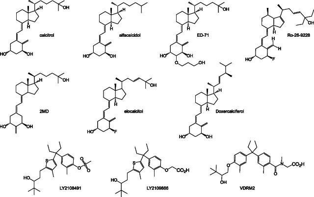
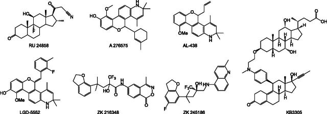
Nuclear receptors are ligand-activated transcription factors and include the receptors for steroid hormones, lipophilic vitamins, sterols, and bile acids. These receptors serve as targets for development of myriad drugs that target a range of disorders. Classically defined ligands that bind to the ligand-binding domain of nuclear receptors, whether they are endogenous or synthetic, either activate receptor activity (agonists) or block activation (antagonists) and due to the ability to alter activity of the receptors are often termed receptor "modulators." The complex pharmacology of nuclear receptors has provided a class of ligands distinct from these simple modulators where ligands display agonist/partial agonist/antagonist function in a tissue or gene selective manner. This class of ligands is defined as selective modulators. Here, we review the development and pharmacology of a range of selective nuclear receptor modulators.


Figures

Fig. 1.
Fig. 1.
Nuclear receptor domain structure and mechanism of action. (A) Nuclear receptors display a conserved modulator domain architecture with an N-terminal AF-1 region (A/B region), followed by zinc-finger DBD (C region), a hinge domain (D region), an LBD containing the AF-2 region (E region), and some receptors have a C-terminal F domain. (B) Mechanistically, nuclear receptors are regulated by small molecule ligands, which generally stabilize the receptor into a conformation suitable to bind coregulator proteins (coactivators or corepressors). Ligands can also modulate posttranslational modification of the receptor. Ultimately, these events have an impact on the expression of receptor-specific target genes by modulating coregulator recruitment at specific DNA-response element sites in the target gene promoter. (C) Schematic illustrating the principle of selective receptor modulation.
Fig. 2.
Fig. 2.
Coactivator binding surface in the LBD composed of a surface formed at the intersection of several structural elements in the LBD, including helices 3, 4, and 12. This surface is called the AF-2 site. Coactivator regions containing a canonical LXXLL motif bind to this surface, docking the three hydrophobic leucine side chains (show in yellow) into a hydrophobic groove. Two key residues in the LBD form a “charge clamp” that helps stabilize this interaction, including a Lysine residue on helix 3 and a Glutamic acid residue on helix 12, which make interactions with the backbone of the LXXLL motif peptide.
Fig. 3.
Fig. 3.
Corepressor binding surface in the LBD. This surface is similar to the coactivator-binding surface but uses a conserved LXXIIXXXI motif to interact with the LBD. The conserved hydrophobic residues (shown in yellow) bind in a hydrophobic groove formed the intersection of helices 3 and 4, but this motif does not engage helix 12 via the “charge clamp.”
Fig. 4.
Fig. 4.
Agonist and antagonist LBD conformations observed in ER crystal structures. Crystal structures of the ER LBD have suggested structural features contributing to ligand-induced agonism and antagonism. (A) The natural agonist, 17β-estradiol, docks in the LBP and positions helix 12 into a conformation referred to as the agonist or active conformation. This conformation forms the coactivator-binding surface as described in Fig. 2. (B) In contrast, when raloxifene is bound in the LBP, the position of helix 12 is rotated with respect to the agonist conformation such that it binds in the AF-2 coactivator-binding surface and thus blocks binding of coactivators via the LXXLL motif. This is referred to as the antagonist, repressive, or inactive conformation. (C) Other ligands such as ICI 182,780 can physically block the AF-2 coactivator-binding surface and do not stabilize helix 12; thus, these ligands are termed pure antagonists. In these panels, helix 12 is blue, and ligands are black.
Fig. 5.
Fig. 5.
Intact structure of PPARγ/RXRα complex on DNA. At the time of this review, only one crystal structure has been reported of an intact nuclear receptor complex: PPARγ/RXRα bound to DNA, ligands, and coactivator peptides. In this structure, the receptors form an asymmetric complex where the LBD of PPARγ contacts not only the LBD of RXRα through the canonical helix 11 heterodimerization surface but also contacts the RXRα DBD, suggesting a feature that could allow molecular communication between the LBD (the site of ligand binding) and DBD (the site of DNA binding). The N-terminal AF-1 region of the receptors was not captured in this complex due to its highly dynamic properties. In this figure, PPARγ is orange, RXRα is blue, ligands are green, and DNA is white.
Fig. 6.
Fig. 6.
HDX study of intact VDR/RXRα complex on DNA. HDX is a powerful technique to monitor the effect of ligand and DNA binding on the dynamics of intact receptor complexes. HDX studies on the intact VDR/RXRα complex were the first to show that binding of the heterodimer to DNA can allosterically permeate changes in receptor dynamics, not only in the DBD but extending to the AF-2 regions in the LBD of the receptors. In this figure, regions colored blue show protection from HDX upon binding DNA, whereas regions colored yellow show increased HDX upon binding DNA. Ligand binding to the LBD was also observed to affect the dynamics of the DBD (not shown).
Fig. 7.
Fig. 7.
ER modulators.
Fig. 8.
Fig. 8.
VDR modulators.
Fig. 9.
Fig. 9.
TR modulators.
Fig. 10.
Fig. 10.
AR modulators.
Fig. 11.
Fig. 11.
GR modulators.
Fig. 12.
Fig. 12.
PPARγ modulators.
Fig. 13.
Fig. 13.
LXR modulators.
Fig. 14.
Fig. 14.
PR modulators.

References

    1. Acton JJ, 3rd, Akiyama TE, Chang CH, Colwell L, Debenham S, Doebber T, Einstein M, Liu K, McCann ME, Moller DE, et al. (2009) Discovery of (2R)-2-(3-3-[(4-methoxyphenyl)carbonyl]-2-methyl-6-(trifluoromethoxy)-1H-indol-1-ylphenoxy)butanoic acid (MK-0533): a novel selective peroxisome proliferator-activated receptor gamma modulator for the treatment of type 2 diabetes mellitus with a reduced potential to increase plasma and extracellular fluid volume. J Med Chem 52:3846–3854. - PubMed
    1. Acton JJ, 3rd, Black RM, Jones AB, Moller DE, Colwell L, Doebber TW, Macnaul KL, Berger J, Wood HB. (2005) Benzoyl 2-methyl indoles as selective PPARγ modulators. Bioorg Med Chem Lett 15:357–362. - PubMed
    1. Adams M, Reginato MJ, Shao DL, Lazar MA, Chatterjee VK. (1997) Transcriptional activation by peroxisome proliferator-activated receptor gamma is inhibited by phosphorylation at a consensus mitogen-activated protein kinase site. J Biol Chem 272:5128–5132. - PubMed
    1. Addo S, Yates RA, Laight A. (2002) A phase I trial to assess the pharmacology of the new oestrogen receptor antagonist fulvestrant on the endometrium in healthy postmenopausal volunteers. Br J Cancer 87:1354–1359. - PMC - PubMed
    1. Adorini L. (2002) 1,25-Dihydroxyvitamin D3 analogs as potential therapies in transplantation. Curr Opin Investig Drugs 3:1458–1463. - PubMed