Review
. 2013 Mar 1;5(3):a008946.
doi: 10.1101/cshperspect.a008946.

The insulin receptor: both a prototypical and atypical receptor tyrosine kinase

Affiliations
Review

The insulin receptor: both a prototypical and atypical receptor tyrosine kinase

Stevan R Hubbard. Cold Spring Harb Perspect Biol. .

Abstract

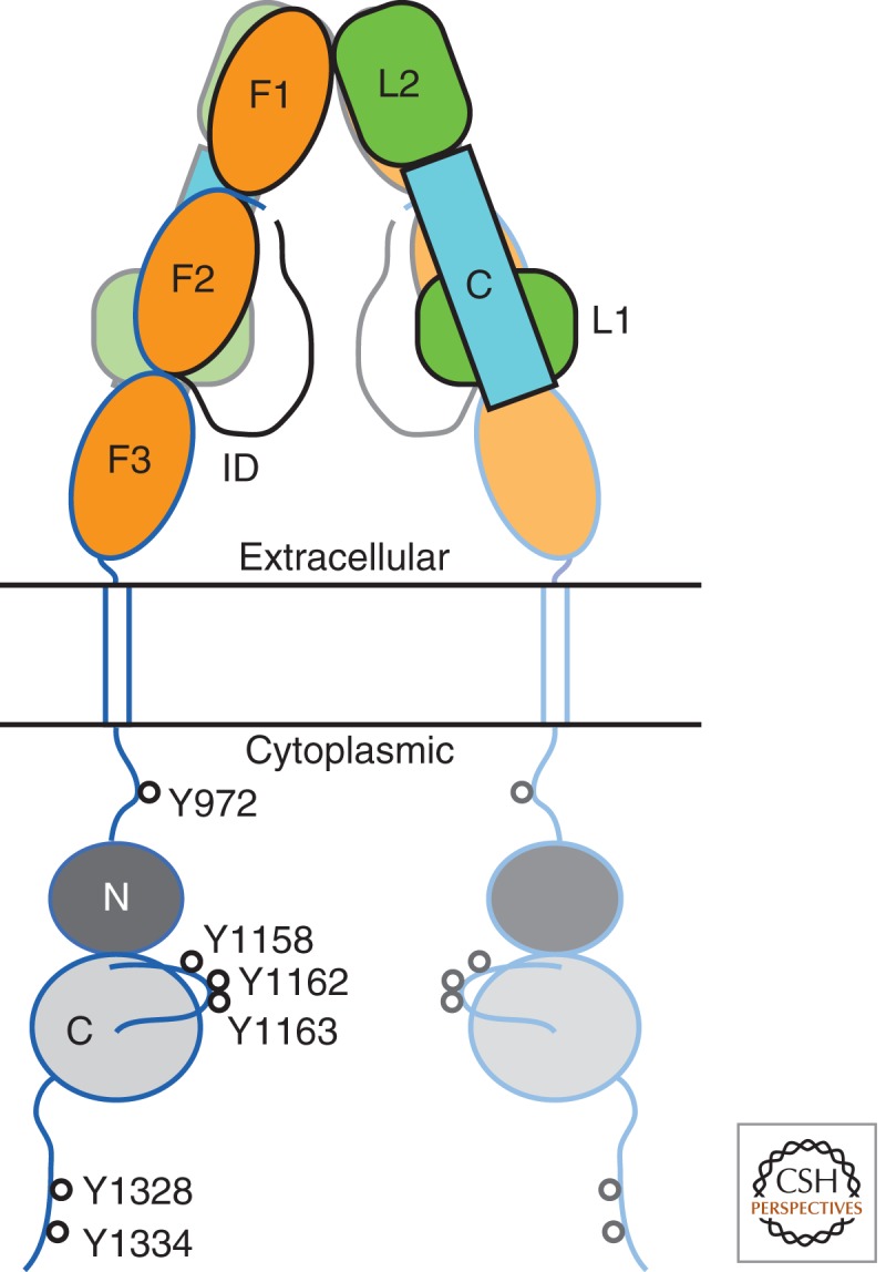
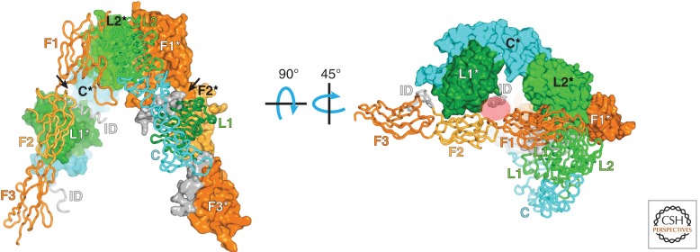
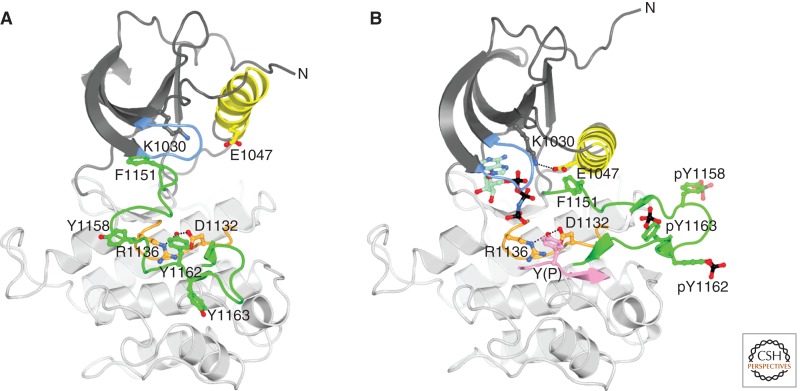
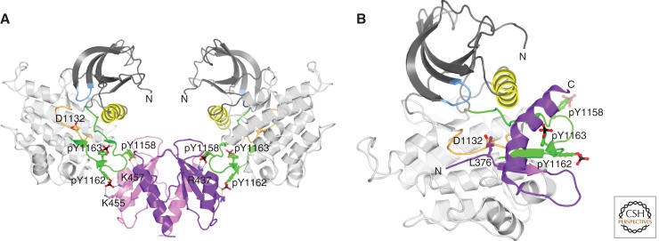
Unlike prototypical receptor tyrosine kinases (RTKs), which are single-chain polypeptides, the insulin receptor (InsR) is a preformed, covalently linked tetramer with two extracellular α subunits and two membrane-spanning, tyrosine kinase-containing β subunits. A single molecule of insulin binds asymmetrically to the ectodomain, triggering a conformational change that is transmitted to the cytoplasmic kinase domains, which facilitates their trans-phosphorylation. As in prototypical RTKs, tyrosine phosphorylation in the juxtamembrane region of InsR creates recruitment sites for downstream signaling proteins (IRS [InsR substrate] proteins, Shc) containing a phosphotyrosine-binding (PTB) domain, and tyrosine phosphorylation in the kinase activation loop stimulates InsR's catalytic activity. For InsR, phosphorylation of the activation loop, which contains three tyrosine residues, also creates docking sites for adaptor proteins (Grb10/14, SH2B2) that possess specialized Src homology-2 (SH2) domains, which are dimeric and engage two phosphotyrosines in the activation loop.


Figures

Figure 1.
Figure 1.
Structure-based schematic diagram of InsR. The α subunits (extracellular, denoted in black outline) comprise two leucine-rich domains (L1 and L2) with an intervening cysteine-rich domain (C), one intact fibronectin type III (FnIII) domain (F1), a partial FnIII domain (F2), and a long insert domain (ID) that contains the site of furin cleavage (L1-C-L2-F1-F2-ID). The β subunits (extracellular and cytoplasmic, denoted in blue outline) contain the completion of the second FnIII domain (F2), followed by an intact FnIII domain (F3), a transmembrane helix, and a cytoplasmic tyrosine kinase domain (N and C lobes). The second αβ half-receptor is shown semitransparent. The major sites of tyrosine autophosphorylation in the cytoplasmic domain are indicated (InsR-B isoform numbering). The molecular twofold axis is vertical, in the plane of the figure.
Figure 2.
Figure 2.
Structure of the ectodomain of InsR. In this disulfide-linked symmetric dimer (apo—no insulin) (McKern et al. 2006; Smith et al. 2010), one of the αβ half-receptors (in front) is shown as a Cα trace, and the other αβ half-receptor (in back) is shown with a molecular surface. The molecular twofold axis is vertical, in the plane of the figure. The two L domains are labeled L1 and L2, the cysteine-rich domain is labeled C, the FnIII domains are labeled F1–3, and the insert domain (end of the α subunit, beginning of the β subunit) is labeled ID. An asterisk in the label denotes a domain from the second half-receptor. The arrows point to the two (identical) insulin-binding sites (although binding of a single insulin molecule is sufficient for receptor activation). In the right panel, the structure has been rotated first by 90°, then by 45°, as indicated. The red oval shows where insulin is thought to bind (corresponds to the arrow on the left in the left panel).
Figure 3.
Figure 3.
Structure of the tyrosine kinase domain of InsR. (A) Ribbon diagram of inactive, unphosphorylated IRK (Hubbard et al. 1994). The N lobe is colored dark gray except for αC and the nucleotide binding loop (blue), and the C lobe is colored light gray except for the catalytic loop (orange) and activation loop (green). Carbon atoms are colored according to their position in the structure, oxygen atoms are colored red, and nitrogen atoms are colored blue. Select side chains are shown in ball-and-stick representation. Hydrogen bonds between Y1162 (the “pseudosubstrate”) in the activation loop and D1132 and R1136 in the catalytic loop are shown as black dashed lines. The amino terminus is labeled (N) (the carboxyl terminus is hidden behind the structure). (B) Ribbon diagram of active, tris-phosphorylated IRK in complex with an ATP analog (AMPPNP) and a peptide substrate (Hubbard 1997). Same coloring as in (A) and, in addition, phosphorus atoms are colored black, carbon atoms of AMPPNP are colored pale green, and the peptide substrate is colored pink. F1151, at the beginning of the activation loop, is part of the DFG motif. Hydrogen bonds between the substrate tyrosine (Y[P]) and D1132 and R1136 are shown by black dashed lines as is the salt bridge between conserved residues K1030 (β3) and E1047 (αC).
Figure 4.
Figure 4.
Structures of InsR-interacting proteins. (A) Ribbon diagram of the dimeric SH2 domain of SH2B2 bound to two IRK (InsR tyrosine kinase domain) molecules (Hu et al. 2003). The two SH2-domain protomers are colored pink and purple. Tris-phosphorylated IRK is colored as in Figure 3B. The molecular twofold axis is vertical. D1132 (catalytic loop), pY1158, pY1162, and pY1163 (activation loop) of IRK are shown in ball-and-stick representation, as are R437, K455, and K457 of SH2B2. Salt bridges are shown by black dashed lines. (B) Ribbon diagram of the BPS region of Grb14 bound to tris-phosphorylated IRK (Depetris et al. 2005). Grb14 BPS is colored purple and the pseudosubstrate residue L376 is shown in ball-and-stick representation. The amino and carboxyl termini of the BPS region are labeled N and C.

References

    1. Artim SC, Mendrola JM, Lemmon MA 2012. Assessing the range of kinase autoinhibition mechanisms in the insulin receptor family. Biochem J 448: 213–220 - PMC - PubMed
    1. Backer JM, Shoelson SE, Weiss MA, Hua QX, Cheatham RB, Haring E, Cahill DC, White MF 1992. The insulin receptor juxtamembrane region contains two independent tyrosine/β-turn internalization signals. J Cell Biol 118: 831–839 - PMC - PubMed
    1. Bandyopadhyay D, Kusari A, Kenner KA, Liu F, Chernoff J, Gustafson TA, Kusari J 1997. Protein-tyrosine phosphatase 1B complexes with the insulin receptor in vivo and is tyrosine-phosphorylated in the presence of insulin. J Biol Chem 272: 1639–1645 - PubMed
    1. Bereziat V, Kasus-Jacobi A, Perdereau D, Cariou B, Girard J, Burnol AF 2002. Inhibition of insulin receptor catalytic activity by the molecular adapter Grb14. J Biol Chem 277: 4845–4852 - PubMed
    1. Bertrand T, Kothe M, Liu J, Dupuy A, Rak A, Berne PF, Davis S, Gladysheva T, Valtre C, Crenne JY, et al. 2012. The crystal structures of TrkA and TrkB suggest key regions for achieving selective inhibition. J Mol Biol 423: 439–453 - PubMed

Publication types