Review
. 2013 Mar 1;5(3):a012591.
doi: 10.1101/cshperspect.a012591.

Prokaryotic nucleotide excision repair

Affiliations
Review

Prokaryotic nucleotide excision repair

Caroline Kisker et al. Cold Spring Harb Perspect Biol. .

Abstract

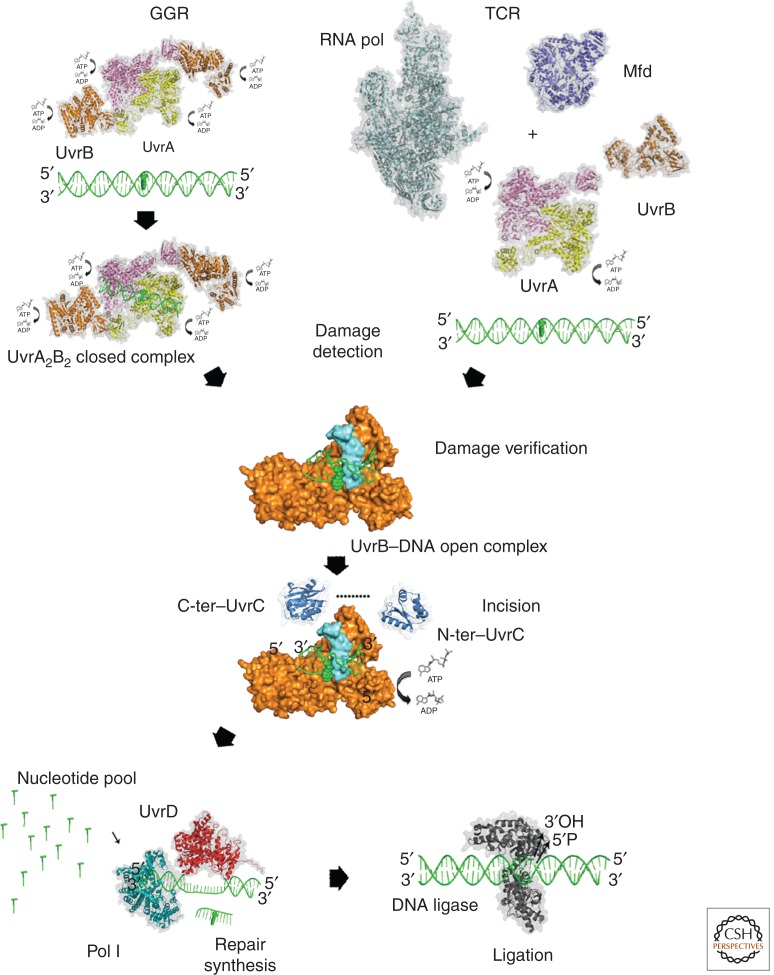
Nucleotide excision repair (NER) has allowed bacteria to flourish in many different niches around the globe that inflict harsh environmental damage to their genetic material. NER is remarkable because of its diverse substrate repertoire, which differs greatly in chemical composition and structure. Recent advances in structural biology and single-molecule studies have given great insight into the structure and function of NER components. This ensemble of proteins orchestrates faithful removal of toxic DNA lesions through a multistep process. The damaged nucleotide is recognized by dynamic probing of the DNA structure that is then verified and marked for dual incisions followed by excision of the damage and surrounding nucleotides. The opposite DNA strand serves as a template for repair, which is completed after resynthesis and ligation.


Figures

Figure 1.
Figure 1.
Schematic representation of the prokaryotic NER pathways. In global genome repair (GGR), the genome is scanned by the heterotetrameric UvrA2–UvrB2 complex in search for damaged nucleotides causing large conformational changes. In transcription-coupled repair, the repair process is initiated by a stalled RNA polymerase on an actively transcribed gene through the interaction of the RNA polymerase with Mfd, which recruits the UvrA dimer or a UvrA2–UvrB heterotrimer to the site of the lesion. Both mechanisms converge into the same pathway and proceed with damage verification by UvrB followed by 3′ and 5′ incisions catalyzed through UvrC. The helicase activity of UvrD is required for the removal of UvrC. The incised strand is excised and repair is completed after the repair patch is synthesized by DNA polymerase I and DNA ligase seals the nick. All protein structures in this figure, with the exception of UvrB, are shown with a transparent surface and in ribbon presentation. UvrB is shown with its surface in orange for domains 1 to 3, and the β-hairpin is shown in cyan. C-ter, Carboxy terminal; N-ter, amino terminal.
Figure 2.
Figure 2.
(Top) Different UvrA domains are shown schematically in the linear polypeptide using the same color codes as are in the ribbon presentations for the different domains within UvrA. (Left) Enlargement of the DNA-binding region of UvrA (PDB entry 2R6F) from B. stearothermophilus. (Center) Superposition of the apo- and DNA-bound UvrA structures. For clarity, two of the monomers are shown as Cα-trace in gray and red, whereas the other two are shown in different shades of gray and in ribbon presentation. (Right) The UvrA2–DNA complex from Thermotoga maritima (PDB entry 3PIH). The color code is the same throughout the figure with the exception of the superposition, and the DNA is shown in orange for the backbone and blue-green spokes for the bases. (Bottom) Structure of the UvrA2–UvrB2 complex (PDB entry 3UWX). The two UvrB molecules are shown in gold.
Figure 3.
Figure 3.
(Top) The UvrB structure is shown schematically within the linear polypeptide. In addition to the colors that match the color coding of the different domains in the structure below, the location of the helicase motifs is indicated by small orange bars. (Left) Overall structure of UvrB in the presence of DNA (PDB entry 2FDC). (Center) The dimeric carboxy-terminal domain of UvrB is shown in ribbon presentation in gray. (Right) Enlargement of the region of the β-hairpin. Residues Tyr-92, Tyr-93, Tyr-95, and Tyr-96 are shown in stick presentation.
Figure 4.
Figure 4.
(Top) Schematic representation of UvrC. No complete structure of UvrC has been solved so far. (Bottom) The two structures from T. maritima represent the 3′ catalytic GIY-YIG domain (PDB entry 1YCZ) and the 5′ RNase H-like catalytic domain as well as the very carboxy-terminal HhH motif (PDB entry 2NRT).
Figure 5.
Figure 5.
Schematic representation of UvrD from E. coli. The two RecA-like domains 1A and 2A are shown in yellow and red, respectively, and the inserted domains 1B and 2B in cyan and green. The DNA is shown in gray (PDB entry 2IS2). In the sequence above, the helicase motifs are indicated by orange bars.
Figure 6.
Figure 6.
Mfd: The seven domains of the TRCF from E. coli, shown schematically (top) and in ribbon presentation (bottom) (PDB entry 2EYQ). The UvrB homology module is indicated in different shades of blue, and the remaining domains are displayed in orange, violet, yellow, green, and red. In the schematic presentation of the polypeptide, the helicase motifs are shown in small orange bars and the TRG is shown in gray.

References

    1. Ahn B 2000. A physical interaction of UvrD with nucleotide excision repair protein UvrB. Mol Cells 10: 592–597 - PubMed
    1. Alexandrovich A, Czisch M, Frenkiel TA, Kelly GP, Goosen N, Moolenaar GF, Chowdhry BZ, Sanderson MR, Lane AN 2001. Solution structure, hydrodynamics and thermodynamics of the UvrB C-terminal domain. J Biomol Struct Dyn 19: 219–236 - PubMed
    1. Aravind L, Walker DR, Koonin EV 1999. Conserved domains in DNA repair proteins and evolution of repair systems. Nucleic Acids Res 27: 1223–1242 - PMC - PubMed
    1. Assenmacher N, Wenig K, Lammens A, Hopfner KP 2006. Structural basis for transcription-coupled repair: The N terminus of Mfd resembles UvrB with degenerate ATPase motifs. J Mol Biol 355: 675–683 - PubMed
    1. Atkinson J, Guy CP, Cadman CJ, Moolenaar GF, Goosen N, McGlynn P 2009. Stimulation of UvrD helicase by UvrAB. J Biol Chem 284: 9612–9623 - PMC - PubMed

Publication types

MeSH terms

LinkOut - more resources