

Review
doi: 10.1101/cshperspect.a012708.
Translesion DNA synthesis and mutagenesis in eukaryotes
Affiliations
- PMID: 23457261
- PMCID: PMC3578355
- DOI: 10.1101/cshperspect.a012708
Item in Clipboard
Review
Translesion DNA synthesis and mutagenesis in eukaryotes
Cold Spring Harb Perspect Biol.
.
Abstract
The structural features that enable replicative DNA polymerases to synthesize DNA rapidly and accurately also limit their ability to copy damaged DNA. Direct replication of DNA damage is termed translesion synthesis (TLS), a mechanism conserved from bacteria to mammals and executed by an array of specialized DNA polymerases. This chapter examines how these translesion polymerases replicate damaged DNA and how they are regulated to balance their ability to replicate DNA lesions with the risk of undesirable mutagenesis. It also discusses how TLS is co-opted to increase the diversity of the immunoglobulin gene hypermutation and the contribution it makes to the mutations that sculpt the genome of cancer cells.
Figures
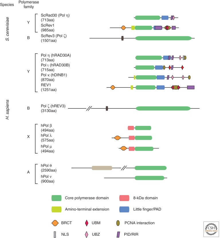
Domain structure of TLS polymerases in S. cerevisiae and Homo sapiens. The diagrams are to scale except that hREV3 and hPol θ are truncated, as indicated by parallel diagonal lines. The REV7 subunit of Pol ζ is not shown. PAD, polymerase-associated domain (also known as the little finger); BRCT, a domain with homology to the BRCA1 carboxyl terminus; UBM and UBZ, ubiquitin-binding domains; PID, polymerase-interacting domain (of REV1); RIR, REV1-interacting region (of other Y-family polymerases).
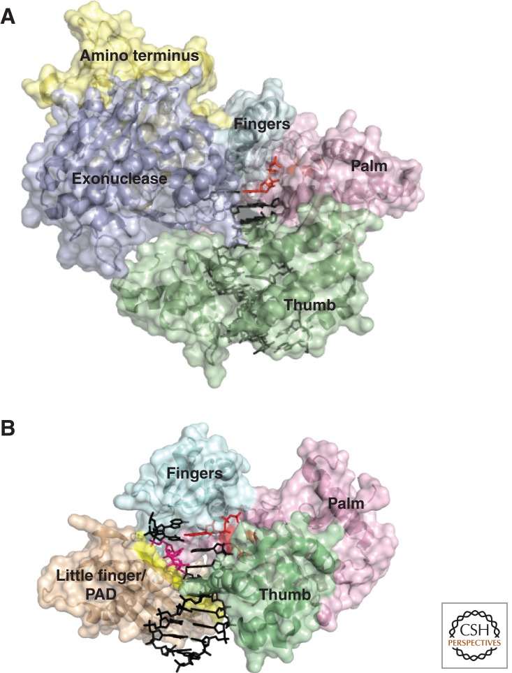
Comparative anatomy of a replicative polymerase and a TLS polymerase. (A) S. cerevisiae polymerase δ, PDB 3IAY (Swan et al. 2009), a replicative B-family polymerase. The domains are shaded: palm, pink; thumb, green; fingers, cyan; exonuclease, purple. The DNA is in black, and the active site in the palm and incoming nucleotide triphosphate is in red. (B) H. sapiens polymerase η, PDB 3SI8 (Biertumpfel et al. 2010), a Y-family TLS polymerase with a T-T CPD in the +2 position. The domains in common with Pol δ are shaded the same. The little-finger domain/PAD is shaded in light brown. The DNA is in black, except the CPD, which is pink. The active site and incoming nucleotide triphosphate pairing with the first base after the CPD are in red. The β-strand splint in the little finger/PAD domain that constrains the CPD is highlighted in yellow.
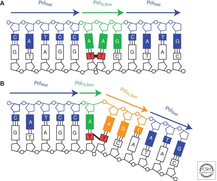
One-step versus two-step TLS. (A) The tracts of DNA synthesized during one-step bypass of a T-T CPD. PolRep, the replicative polymerases Pol δ and Pol ε. In the case illustrated, bypass of a CPD, the inserter and extender polymerase (PolTLSins/ext) is Pol η, which can replicate this lesion on its own, aided by its large active site pocket and molecular split (see text). The extent of its synthesis patch is limited either by its dissociation from the DNA induced by a steric clash between the lesion and the little-finger domain as the chain elongates or by the primer at the end of a single-stranded gap. (B) The tracts of DNA synthesized during two-step bypass of a T-T (6-4) photoproduct. Like many lesions, the highly distorting (6-4) photoproduct is not bypassed in a single step. Instead, an inserter polymerase (PolTLSins) incorporates a base opposite the 3′T of the lesion, but cannot extend opposite the 5′T. Instead, this is carried out by an extender polymerase (PolTLSext), most commonly Pol ζ, which can deal with the highly misaligned terminus.
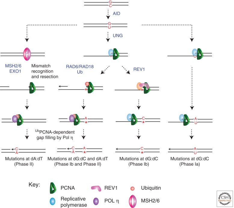
The role of TLS in Ig gene somatic hypermutation. Ig gene hypermutation is initiated by AID, which deaminates dC in the Ig variable-region genes to produce uracil. This triggers a number of mutagenic consequences. In the right column, U is replicated by Pol δ or Pol ε, resulting in C/G to T/A transitions (Phase Ia [Neuberger et al. 2003]). Alternatively, U is excised to generate an abasic site (central columns). This can be replicated by a polymerase capable of TLS to generate all possible mutations at dC (Phase Ib). REV1 is illustrated and is responsible for C/G to G/C transversions. Alternatively, replication of the abasic site stimulates PCNA ubiquitination, possibly by gap formation following replication arrest, and this recruits Pol η. This can result in mutations at the site of dC deamination to generate Phase Ib mutation and could also lead to Phase II mutations at A/T base pairs generated by misincorporation on undamaged DNA by Pol η. Finally, the initial U/G mismatch can be recognized as a mismatch by MSH2/6, resulting in resection by EXO1 and formation of a gap. This, in turn, stimulates PCNA ubiquitination, recruitment of Pol η, and Phase II mutagenesis at A/T base pairs.
References
-
- Albertella MR, Green CM, Lehmann AR, O’Connor MJ 2005a. A role for polymerase η in the cellular tolerance to cisplatin-induced damage. Cancer Res 65: 9799–9806 - PubMed
-
- Albertella MR, Lau A, O’Connor MJ 2005b. The overexpression of specialized DNA polymerases in cancer. DNA Repair (Amst) 4: 583–593 - PubMed
Publication types
MeSH terms
Substances
Grants and funding
LinkOut - more resources
Full Text Sources
Other Literature Sources