. 2013;8(2):e55075.
doi: 10.1371/journal.pone.0055075. Epub 2013 Feb 15.

The anti-migratory effects of FKBPL and its peptide derivative, AD-01: regulation of CD44 and the cytoskeletal pathway

Affiliations

The anti-migratory effects of FKBPL and its peptide derivative, AD-01: regulation of CD44 and the cytoskeletal pathway

Anita Yakkundi et al. PLoS One. 2013.

Abstract

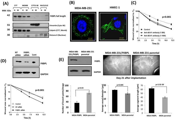
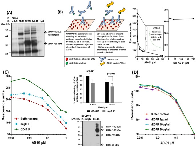
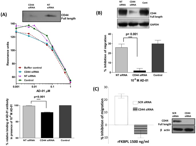
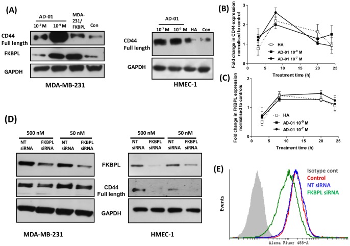
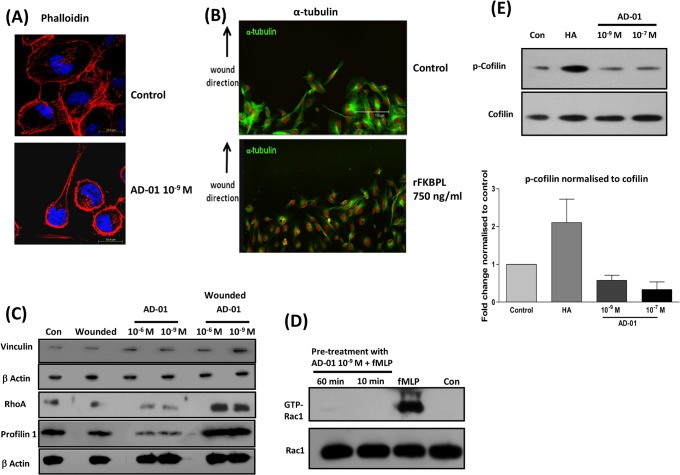
FK506 binding protein-like (FKBPL) and its peptide derivatives exert potent anti-angiogenic activity in vitro and in vivo and control tumour growth in xenograft models, when administered exogenously. However, the role of endogenous FKBPL in angiogenesis is not well characterised. Here we investigated the molecular effects of the endogenous protein and its peptide derivative, AD-01, leading to their anti-migratory activity. Inhibition of secreted FKBPL using a blocking antibody or siRNA-mediated knockdown of FKBPL accelerated the migration of human microvascular endothelial cells (HMEC-1). Furthermore, MDA-MB-231 tumour cells stably overexpressing FKBPL inhibited tumour vascular development in vivo suggesting that FKBPL secreted from tumour cells could inhibit angiogenesis. Whilst FKBPL and AD-01 target CD44, the nature of this interaction is not known and here we have further interrogated this aspect. We have demonstrated that FKBPL and AD-01 bind to the CD44 receptor and inhibit tumour cell migration in a CD44 dependant manner; CD44 knockdown abrogated AD-01 binding as well as its anti-migratory activity. Interestingly, FKBPL overexpression and knockdown or treatment with AD-01, regulated CD44 expression, suggesting a co-regulatory pathway for these two proteins. Downstream of CD44, alterations in the actin cytoskeleton, indicated by intense cortical actin staining and a lack of cell spreading and communication were observed following treatment with AD-01, explaining the anti-migratory phenotype. Concomitantly, AD-01 inhibited Rac-1 activity, up-regulated RhoA and the actin binding proteins, profilin and vinculin. Thus the anti-angiogenic protein, FKBPL, and AD-01, offer a promising and alternative approach for targeting both CD44 positive tumours and vasculature networks.


Conflict of interest statement

Competing Interests: The authors have received financial support from BBSRC and Almac Discovery, Northern Ireland for consumables and salaries of authors AY, L. McCallum, HD at various times. Furthermore TR and DGH are patent holders on: Robson T, Hirst GH, Valentine A, O'Rourke M. FKBP-L AND USES THEREOF. PCT/GB2007/002107 (WO/2007/141533). AU, CA, EP, JP, NZ, US. 2007. This patent was licensed to Almac Discovery from QUB in 2006. The authors declare that this does not alter their adherence to PLOS ONE's policies on sharing data and materials.

Figures

Figure 1
Figure 1. FKBPL is present in various cell compartments and regulates cell migration and tumour vasculature.
(A) Representative blot demonstrating that FKBPL is present predominantly in the cytosol and membrane compartments of both HMEC-1 (H) and MDA-MB-231 (M) cells and in the nuclear fraction of MDA-MB-231 cells. Protein extracts from each subcellular compartment probed with specific compartmental markers, vimentin, calpain and histone-H1 were used as loading controls. (B) Representative confocal images (60x) of MDA-MB-231 and HMEC-1 cells fixed, permeabilised and stained with DAPI (blue) and with anti-AD-01 primary antibody and Alexa-488 tagged secondary antibody demonstrating vesicular staining for FKBPL (green); n = 3. (C) Anti-AD-01 antibody targets the active domain of FKBPL and accelerates HMEC-1 cell migration in comparison to cells treated with an isotype control. Data points show means ± SEM; n = 3 (D) FKBPL knockdown with siRNA accelerated migration of HMEC-1 cells in comparison to un-transfected and NT-siRNA-transfected cells. Data points show means ± SEM; n = 3. Cell migration was assessed using scratch wound assay. Wound size is normalised to that of T0. p-value was determined using two-way ANOVA. (E) Intravital microscopy images (20x) representing disruption of tumour vasculature in vivo in FKBPL-overexpressing MDA-MB-231 xenografts in comparison to those derived from parental MDA-MB-231 cells. Tumours (21 days) were imaged using Epi-fluoresence microscopy following injection of mice with FITC-Dextran. Quantification of vessel dynamics was carried out on 3D images using ImageJ software. n = 5 mice per treatment group (p-value was determined using two-tailed T –test).
Figure 2
Figure 2. FKBPL and its peptide derivative, AD-01, bind CD44.
(A) Representative western blot showing FKBPL co-immunoprecipitated with CD44 in HMEC-1 cells; immuno-blotted with anti-CD44 antibody; n = 3; Cdc42 was used as a positive control for the CD44 interaction and rabbit and murine IgGs were used as negative controls. (B) Schematic diagram of the Biacore assay using AD-01 immobilised on CM5 chip surface. Binding of anti-AD-01 antibody to AD-01-CM5 surface was inhibited by AD-01 in solution in a dose dependent manner, with excellent sensitivity in lower concentration range of peptide; 1–500 nM. Scrambled AD-01, used as a negative control, did not demonstrate any binding to anti-AD-01 up to 200 µM. Competition of the anti-AD-01 antibody interaction with its cellular partner/s results in increased binding of anti-AD-01 antibody on chip surface. (C) Representative graph demonstrating that AD-01 specifically binds to CD44 immunoprecipitated from MDA-MB-231 cells using the assay described. CD44 was immuno-purified from cell lysate and analysed using Biacore Q. Isotype control mIgG antibody was used as control. Bar charts show the relative binding of anti-AD-01 antibody in presence of AD-01, calculated as the percentage of the maximum resonance binding units in the presence of 0.001 and 0.01 µM AD-01. Data points show means ± SEM of 5 independent experiments (p-value was determined by one way ANOVA). (D) No competition of anti-AD-01 antibody binding to immobilised AD-01 was obtained in the presence of various concentrations of rEGFR indicating a specificity of AD-01-CD44 interaction.
Figure 3
Figure 3. Binding and anti-migratory activity of AD-01 is dependent on CD44.
(A) Representative graph demonstrating extracts of MDA-MB-231 un-transfected control/NT siRNA transfected cells render increased binding of anti-AD-01 antibody to AD-01 immobilised on CM5 chip surface using the specific binding assay described in Fig. 2. This binding was abrogated in lysates derived from CD44 knockdown cells. Buffer control comprised of HBS buffer with indicated amounts of AD-01. Bar charts demonstrate significant abrogation of this binding in the presence of 10−9 M AD-01 in lysates from cells transfected with CD44 siRNA. Data points show means ± SEM; n = 4. (B) Treatment with AD-01 at 10−9 M results in inhibition of migration in un-transfected and NT siRNA transfected MDA-MB-231 cells. This inhibition of migration was abrogated upon CD44 knockdown using targeted siRNA. Data points show means ± SEM; n = 5. (p-value was determined using one-way ANOVA). (C) Anti-migratory effect of rFKBPL 1500 ng/ml was abrogated in PC3 cells transfected with CD44 targeted siRNA in comparison to those transfected with scrambled siRNA; n = 3.
Figure 4
Figure 4. AD-01 and FKBPL regulate CD44 expression.
(A) Treatment with AD-01 up-regulated CD44 expression. Representative blots of cell lysates from MDA-MB-231/HMEC-1 treated with AD-01 or HA (0.1 mg/ml) for 24 h and MDA-MB-231/FKBPL overexpressing cells, probed with anti-CD44, FKBPL or GAPDH antibodies (n = 3). Transcriptional regulation of CD44 (B) and FKBPL (C) upon treatment with AD-01/HA was assessed using quantitative RT-PCR. Data points show mean fold change ± SEM in FKBPL or CD44 expression in cDNA prepared from cells treated with AD-01 in comparison to control untreated cells; n = 3. (D) Representative western blots demonstrating dose dependent down-regulation of FKBPL upon transfection with FKBPL targeted siRNA for 72 h, resulting in a corresponding down-regulation of CD44 protein; n = 3. (E) Flow cytometric analysis of cell surface expression of CD44 in MDA-MB-231 cells transfected with FKBPL siRNA in comparison to NT siRNA and stained with anti-CD44 and Alexa-488 conjugated secondary antibody; n = 3.
Figure 5
Figure 5. AD-01 and FKBPL mediate cytoskeletal changes
and AD-01 disrupts the RhoA-Rac1 dynamics. (A) Confocal images (60x) representing the changes in phalloidin/F-actin dynamics in MDA-MB-231 upon wounding and treatment with AD-01 (10−9 M) for 24 h; n = 3. Treated monolayers were fixed and stained with TRITC-phallodin (red) and DAPI (blue). Intense actin staining was accompanied by the loss in cell direction and communication. (B) Images (40x) representing disruption in tubulin distribution in HMEC-1 cell monolayers, wounded and treated with rFKBPL 750 ng/ml for 5 h. Fixed monolayers were stained for tubulin, followed by FITC conjugated secondary antibody (green) and nucleus with PI (red). (C) RhoA expression was increased after wounding and treatment with AD-01 for 24 h, resulting in a concomitant increase in the downstream actin binding proteins vinculin and profilin. Cells monolayers were treated with AD-01 (± wounding) for 24 h and total cell lysates were subjected to immuno blotting as indicated. (D) Treatment with AD-01 (10−9 M) for 10/60 min inhibited fMLP induction (30 sec) of GTP-Rac-1 in HMEC-1 cells. Cell lysates of treated monolayers were subjected to Rac GTPase pull down assay; n = 3. (E) Representative western blots and quantitative densitometric analysis demonstrating an inhibition of cofilin phosphorylation after treatment with AD-01. HA treatment up-regulated cofilin phosphorylation maintaining its inactive state. HMEC-1 cell monolayers were treated with AD-01 for 3 h or HA for 10 min, and the extracted membrane fractions were subjected to immunoblotting with cofilin/p-cofilin; n = 3.

References

    1. Guidolin D (2010) The multifaceted world of angiogenesis control. Expert Opinion Ther Targets 14: 1135–1138. - PubMed
    1. Cao Y (2010) Angiogenesis: What can it offer for future medicine? Exp Cell Res 316: 1304–1308. - PubMed
    1. Stone EM (2006) A very effective treatment for neovascular macular degeneration. N Engl J Med 355: 1493–1495. - PubMed
    1. Ferrara N, Hillan KJ, Gerber HP, Novotny W (2004) Discovery and development of bevacizumab, an anti-VEGF antibody for treating cancer. Nat Rev Drug Discov 3: 391–400. - PubMed
    1. Motzer RJ, Bukowski RM (2006) Targeted therapy for metastatic renal cell carcinoma. J Clin Oncol 24: 5601–5608. - PubMed

Publication types

MeSH terms